[English] 日本語
 Yorodumi
- PDB-9dz2: Cryo-EM structure of Sudan ebolavirus glycoprotein complexed with... -
+ Open data
- Basic information
| Entry | Database: PDB / ID: 9dz2 | |||||||||
|---|---|---|---|---|---|---|---|---|---|---|
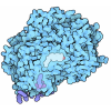
| Title | Cryo-EM structure of Sudan ebolavirus glycoprotein complexed with hNPC1-C | |||||||||
 Components Components |
| |||||||||
 Keywords Keywords | VIRAL PROTEIN / SUDV / hNPC1-C / glycoprotein | |||||||||
| Function / homology |  Function and homology information Function and homology informationcyclodextrin metabolic process / cholesterol storage / membrane raft organization / intracellular cholesterol transport / intracellular lipid transport / sterol transport / intestinal cholesterol absorption / LDL clearance / negative regulation of epithelial cell apoptotic process / cholesterol transfer activity ...cyclodextrin metabolic process / cholesterol storage / membrane raft organization / intracellular cholesterol transport / intracellular lipid transport / sterol transport / intestinal cholesterol absorption / LDL clearance / negative regulation of epithelial cell apoptotic process / cholesterol transfer activity / cholesterol transport / : / programmed cell death / bile acid metabolic process / establishment of protein localization to membrane / adult walking behavior / cholesterol efflux / lysosomal transport / cholesterol binding / cellular response to steroid hormone stimulus / negative regulation of macroautophagy / cellular response to low-density lipoprotein particle stimulus / response to cadmium ion / cholesterol metabolic process / negative regulation of TORC1 signaling / neurogenesis / cholesterol homeostasis / macroautophagy / liver development / autophagy / endocytosis / transmembrane signaling receptor activity / nuclear envelope / late endosome membrane / signaling receptor activity / virus receptor activity / gene expression / clathrin-dependent endocytosis of virus by host cell / symbiont-mediated-mediated suppression of host tetherin activity / entry receptor-mediated virion attachment to host cell / lysosome / symbiont-mediated suppression of host innate immune response / membrane raft / response to xenobiotic stimulus / lysosomal membrane / fusion of virus membrane with host endosome membrane / viral envelope / symbiont entry into host cell / perinuclear region of cytoplasm / host cell plasma membrane / virion membrane / endoplasmic reticulum / Golgi apparatus / extracellular exosome / extracellular region / membrane / plasma membrane Similarity search - Function | |||||||||
| Biological species |  Homo sapiens (human) Homo sapiens (human) Sudan ebolavirus Sudan ebolavirus | |||||||||
| Method | ELECTRON MICROSCOPY / single particle reconstruction / cryo EM / Resolution: 3.31 Å | |||||||||
 Authors Authors | Bu, F. / Ye, G. / Liu, B. / Li, F. | |||||||||
| Funding support |  United States, 2items United States, 2items
| |||||||||
 Citation Citation |  Journal: Commun Biol / Year: 2025 Journal: Commun Biol / Year: 2025Title: Cryo-EM structure of Sudan ebolavirus glycoprotein complexed with its human endosomal receptor NPC1. Authors: Fan Bu / Gang Ye / Hailey Turner-Hubbard / Morgan Herbst / Bin Liu / Fang Li /  Abstract: Sudan ebolavirus (SUDV), like Ebola ebolavirus (EBOV), poses a significant threat to global health and security due to its high lethality. However, unlike EBOV, there are no approved vaccines or ...Sudan ebolavirus (SUDV), like Ebola ebolavirus (EBOV), poses a significant threat to global health and security due to its high lethality. However, unlike EBOV, there are no approved vaccines or treatments for SUDV, and its structural interaction with the endosomal receptor NPC1 remains unclear. This study compares the glycoproteins of SUDV and EBOV (in their proteolytically primed forms) and their binding to human NPC1 (hNPC1). The findings reveal that the SUDV glycoprotein binds significantly more strongly to hNPC1 than the EBOV glycoprotein. Using cryo-EM, we determined the structure of the SUDV glycoprotein/hNPC1 complex, identifying four key residues in the SUDV glycoprotein that differ from those in the EBOV glycoprotein and influence hNPC1 binding: Ile79, Ala141, and Pro148 enhance binding, while Gln142 reduces it. Collectively, these residue differences account for SUDV's stronger binding affinity for hNPC1. This study provides critical insights into receptor recognition across all viruses in the ebolavirus genus, including their interactions with receptors in bats, their suspected reservoir hosts. These findings advance our understanding of ebolavirus cell entry, tissue tropism, and host range. | |||||||||
| History |
|
- Structure visualization
| Structure viewer | Molecule:  Molmil Molmil Jmol/JSmol Jmol/JSmol |
|---|
- Downloads & links
- Download
| PDBx/mmCIF format |  9dz2.cif.gz 9dz2.cif.gz | 241.8 KB | Display |  PDBx/mmCIF format PDBx/mmCIF format |
|---|---|---|---|---|
| PDB format |  pdb9dz2.ent.gz pdb9dz2.ent.gz | 189.7 KB | Display |  PDB format PDB format |
| PDBx/mmJSON format |  9dz2.json.gz 9dz2.json.gz | Tree view |  PDBx/mmJSON format PDBx/mmJSON format | |
| Others |  Other downloads Other downloads |
-Validation report
| Arichive directory |  https://data.pdbj.org/pub/pdb/validation_reports/dz/9dz2 https://data.pdbj.org/pub/pdb/validation_reports/dz/9dz2 ftp://data.pdbj.org/pub/pdb/validation_reports/dz/9dz2 ftp://data.pdbj.org/pub/pdb/validation_reports/dz/9dz2 | HTTPS FTP |
|---|
-Related structure data
| Related structure data |  47323MC M: map data used to model this data C: citing same article ( |
|---|---|
| Similar structure data | Similarity search - Function & homology  F&H Search F&H Search |
- Links
Links
- Assembly
| Deposited unit | 
|
|---|---|
| 1 |
|
- Components
| #1: Protein | Mass: 31792.486 Da / Num. of mol.: 2 Source method: isolated from a genetically manipulated source Source: (gene. exp.)  Homo sapiens (human) / Gene: NPC1 / Production host: Homo sapiens (human) / Gene: NPC1 / Production host:  Homo sapiens (human) / References: UniProt: O15118 Homo sapiens (human) / References: UniProt: O15118#2: Protein | Mass: 21546.752 Da / Num. of mol.: 3 Source method: isolated from a genetically manipulated source Source: (gene. exp.)  Sudan ebolavirus / Gene: GP / Production host: Sudan ebolavirus / Gene: GP / Production host:  Homo sapiens (human) / References: UniProt: Q7T9D9 Homo sapiens (human) / References: UniProt: Q7T9D9#3: Protein | Mass: 18787.199 Da / Num. of mol.: 3 Source method: isolated from a genetically manipulated source Source: (gene. exp.)  Sudan ebolavirus / Gene: GP / Production host: Sudan ebolavirus / Gene: GP / Production host:  Homo sapiens (human) / References: UniProt: Q7T9D9 Homo sapiens (human) / References: UniProt: Q7T9D9Has protein modification | Y | |
|---|
-Experimental details
-Experiment
| Experiment | Method: ELECTRON MICROSCOPY |
|---|---|
| EM experiment | Aggregation state: PARTICLE / 3D reconstruction method: single particle reconstruction |
- Sample preparation
| Component | Name: SUDV GP complexed with NPC1-C / Type: COMPLEX / Entity ID: all / Source: RECOMBINANT |
|---|---|
| Source (natural) | Organism:  Sudan ebolavirus Sudan ebolavirus |
| Source (recombinant) | Organism:  Homo sapiens (human) Homo sapiens (human) |
| Buffer solution | pH: 6 |
| Specimen | Embedding applied: NO / Shadowing applied: NO / Staining applied: NO / Vitrification applied: YES |
| Specimen support | Grid material: COPPER / Grid mesh size: 300 divisions/in. / Grid type: Quantifoil R1.2/1.3 |
| Vitrification | Cryogen name: ETHANE |
- Electron microscopy imaging
| Experimental equipment |  Model: Titan Krios / Image courtesy: FEI Company |
|---|---|
| Microscopy | Model: TFS KRIOS |
| Electron gun | Electron source:  FIELD EMISSION GUN / Accelerating voltage: 300 kV / Illumination mode: FLOOD BEAM FIELD EMISSION GUN / Accelerating voltage: 300 kV / Illumination mode: FLOOD BEAM |
| Electron lens | Mode: BRIGHT FIELD / Nominal defocus max: 2000 nm / Nominal defocus min: 1000 nm |
| Image recording | Electron dose: 53.7 e/Å2 / Film or detector model: GATAN K3 BIOQUANTUM (6k x 4k) |
- Processing
| EM software | Name: PHENIX / Version: 1.21.1_5286: / Category: model refinement | ||||||||||||||||||||||||
|---|---|---|---|---|---|---|---|---|---|---|---|---|---|---|---|---|---|---|---|---|---|---|---|---|---|
| CTF correction | Type: PHASE FLIPPING AND AMPLITUDE CORRECTION | ||||||||||||||||||||||||
| 3D reconstruction | Resolution: 3.31 Å / Resolution method: FSC 0.143 CUT-OFF / Num. of particles: 159976 / Symmetry type: POINT | ||||||||||||||||||||||||
| Atomic model building | PDB-ID: 5f1b Accession code: 5f1b / Source name: PDB / Type: experimental model | ||||||||||||||||||||||||
| Refine LS restraints |
|
 Movie
Movie Controller


 PDBj
PDBj


