 Movie
Movie Controller

[English] 日本語
 Yorodumi
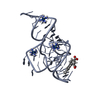
- PDB-9dxl: RhoBAST RNA aptamer in complex with the SpyRho555 analogue, MaP555 -
+ Open data

- Basic information
| Entry | Database: PDB / ID: 9dxl | |||||||||
|---|---|---|---|---|---|---|---|---|---|---|
| Title | RhoBAST RNA aptamer in complex with the SpyRho555 analogue, MaP555 | |||||||||
 Components Components | RhoBAST aptamer | |||||||||
 Keywords Keywords | RNA / aptamer / fluorophore / dye / MaP555 | |||||||||
| Function / homology | : / : / RNA / RNA (> 10) Function and homology information Function and homology information | |||||||||
| Biological species | synthetic construct (others) | |||||||||
| Method |  X-RAY DIFFRACTION / MOLECULAR REPLACEMENT / Resolution: 2.8 Å X-RAY DIFFRACTION / MOLECULAR REPLACEMENT / Resolution: 2.8 Å | |||||||||
 Authors Authors | Siwik, S.H. / Batey, R.T. | |||||||||
| Funding support |  United States, 2items United States, 2items
| |||||||||
 Citation Citation | Journal: Nucleic Acids Res. / Year: 2025 Title: Structural basis for ring-opening fluorescence by the RhoBAST RNA aptamer. Authors: Siwik, S.H. / Wierzba, A.J. / Lennon, S.R. / Olenginski, L.T. / Palmer, A.E. / Batey, R.T. | |||||||||
| History |
|
- Structure visualization
| Structure viewer | Molecule: MolmilJmol/JSmol |
|---|
- Downloads & links
-Download
| PDBx/mmCIF format | 9dxl.cif.gz | 92 KB | Display | PDBx/mmCIF format |
|---|---|---|---|---|
| PDB format | pdb9dxl.ent.gz | Display | PDB format | |
| PDBx/mmJSON format | 9dxl.json.gz | Tree view | PDBx/mmJSON format | |
| Others |  Other downloads Other downloads |
-Validation report
| Arichive directory | https://data.pdbj.org/pub/pdb/validation_reports/dx/9dxlftp://data.pdbj.org/pub/pdb/validation_reports/dx/9dxl | HTTPS FTP |
|---|
-Related structure data
-Links
 PDBj
PDBj


- Assembly
| Deposited unit | 
| ||||||||||||
|---|---|---|---|---|---|---|---|---|---|---|---|---|---|
| 1 | 
| ||||||||||||
| 2 | 
| ||||||||||||
| Unit cell |
|
-Components
| #1: RNA chain | Mass: 15586.393 Da / Num. of mol.: 2 / Source method: obtained synthetically Details: T7 RNA polymerase transcript of an in vitro selected aptamer Source: (synth.) synthetic construct (others) #2: Chemical | ChemComp-BA /   Mass: 137.327 Da / Num. of mol.: 9 / Source method: obtained synthetically / Formula: Ba Mass: 137.327 Da / Num. of mol.: 9 / Source method: obtained synthetically / Formula: Ba#3: Chemical | ChemComp-A1BC9 / | Mass: 537.607 Da / Num. of mol.: 1 / Source method: obtained synthetically / Formula: C27H29N4O6S / Feature type: SUBJECT OF INVESTIGATION #4: Water | ChemComp-HOH / |  Mass: 18.015 Da / Num. of mol.: 34 / Source method: isolated from a natural source / Formula: H2O Mass: 18.015 Da / Num. of mol.: 34 / Source method: isolated from a natural source / Formula: H2OHas ligand of interest | Y | Has protein modification | N | |
|---|
-Experimental details
-Experiment
| Experiment | Method: X-RAY DIFFRACTION / Number of used crystals: 1 |
|---|
- Sample preparation
| Crystal | Density Matthews: 2.88 Å3/Da / Density % sol: 57.4 % / Description: elongated prisms |
|---|---|
| Crystal grow | Temperature: 292 K / Method: vapor diffusion, hanging drop / pH: 6 Details: 40 mM MES, 10 mM barium chloride, 80 mM potassium chloride, 25% MPD, 12 mM spermine tetrahydrochloride |
-Data collection
| Diffraction | Mean temperature: 100 K / Serial crystal experiment: N |
|---|---|
| Diffraction source | Source: SEALED TUBE / Type: RIGAKU MICROMAX-003 / Wavelength: 1.54178 Å |
| Detector | Type: DECTRIS PILATUS 200K / Detector: PIXEL / Date: Sep 7, 2024 |
| Radiation | Monochromator: Ni filter / Protocol: SINGLE WAVELENGTH / Monochromatic (M) / Laue (L): M / Scattering type: x-ray |
| Radiation wavelength | Wavelength: 1.54178 Å / Relative weight: 1 |
| Reflection | Resolution: 2.8→20 Å / Num. obs: 8812 / % possible obs: 98 % / Redundancy: 2.7 % / Rpim(I) all: 0.074 / Rrim(I) all: 0.127 / Χ2: 1.541 / Net I/av σ(I): 11.56 / Net I/σ(I): 9.4 |
| Reflection shell | Resolution: 2.8→2.9 Å / Redundancy: 2.4 % / Mean I/σ(I) obs: 2.69 / Num. unique obs: 839 / CC1/2: 0.815 / CC star: 0.948 / Rpim(I) all: 0.074 / Rrim(I) all: 0.127 / % possible all: 96.2 |
- Processing
| Software |
| |||||||||||||||||||||||||||||||||||||||||||||||||
|---|---|---|---|---|---|---|---|---|---|---|---|---|---|---|---|---|---|---|---|---|---|---|---|---|---|---|---|---|---|---|---|---|---|---|---|---|---|---|---|---|---|---|---|---|---|---|---|---|---|---|
| Refinement | Method to determine structure: MOLECULAR REPLACEMENT / Resolution: 2.8→19.27 Å / Cross valid method: FREE R-VALUE / σ(F): 21.83 / Phase error: 40.8904 Stereochemistry target values: GeoStd + Monomer Library + CDL v1.2
| |||||||||||||||||||||||||||||||||||||||||||||||||
| Solvent computation | Shrinkage radii: 0.9 Å / VDW probe radii: 1.11 Å / Solvent model: FLAT BULK SOLVENT MODEL | |||||||||||||||||||||||||||||||||||||||||||||||||
| Displacement parameters | Biso mean: 73.1 Å2 | |||||||||||||||||||||||||||||||||||||||||||||||||
| Refinement step | Cycle: LAST / Resolution: 2.8→19.27 Å
| |||||||||||||||||||||||||||||||||||||||||||||||||
| Refine LS restraints |
| |||||||||||||||||||||||||||||||||||||||||||||||||
| LS refinement shell |
|
