+ データを開く
- 基本情報
基本情報
| 登録情報 | データベース: PDB / ID: 9dvf | ||||||||||||||||||
|---|---|---|---|---|---|---|---|---|---|---|---|---|---|---|---|---|---|---|---|
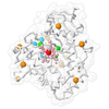
| タイトル | Structure of the native PLP synthase subunit PdxS from Methanosarcina acetivorans | ||||||||||||||||||
 要素 要素 | Pyridoxal 5'-phosphate synthase subunit PdxS | ||||||||||||||||||
 キーワード キーワード | LYASE / pyridoxal 5'-phosphate synthase / TIM barrel / vitamer biosynthesis / BIOSYNTHETIC PROTEIN | ||||||||||||||||||
| 機能・相同性 |  機能・相同性情報 機能・相同性情報amine-lyase activity / pyridoxal 5'-phosphate synthase (glutamine hydrolysing) / pyridoxal 5'-phosphate synthase (glutamine hydrolysing) activity / pyridoxal phosphate biosynthetic process / pyridoxine biosynthetic process / amino acid metabolic process 類似検索 - 分子機能 | ||||||||||||||||||
| 生物種 |  Methanosarcina acetivorans C2A (古細菌) Methanosarcina acetivorans C2A (古細菌) | ||||||||||||||||||
| 手法 | 電子顕微鏡法 / 単粒子再構成法 / クライオ電子顕微鏡法 / 解像度: 3.38 Å | ||||||||||||||||||
 データ登録者 データ登録者 | Agnew, A. / Humm, E. / Zhou, K. / Gunsalus, R. / Zhou, Z.H. | ||||||||||||||||||
| 資金援助 |  米国, 5件 米国, 5件
| ||||||||||||||||||
 引用 引用 |  ジャーナル: mBio / 年: 2025 ジャーナル: mBio / 年: 2025タイトル: Structure and identification of the native PLP synthase complex from lysate. 著者: Angela Agnew / Ethan Humm / Kang Zhou / Robert P Gunsalus / Z Hong Zhou /  要旨: Many protein-protein interactions behave differently in biochemically purified forms as compared to their states. As such, determining native protein structures may elucidate structural states ...Many protein-protein interactions behave differently in biochemically purified forms as compared to their states. As such, determining native protein structures may elucidate structural states previously unknown for even well-characterized proteins. Here, we apply the bottom-up structural proteomics method, , toward a model methanogenic archaeon. While they are keystone organisms in the global carbon cycle and active members of the human microbiome, there is a general lack of characterization of methanogen enzyme structure and function. Through the approach, we successfully reconstructed and identified the native pyridoxal 5'-phosphate (PLP) synthase (PdxS) complex directly from cryogenic electron microscopy (cryo-EM) images of fractionated cellular lysate. We found that the native PdxS complex exists as a homo-dodecamer of PdxS subunits, and the previously proposed supracomplex containing both the synthase (PdxS) and glutaminase (PdxT) was not observed in cellular lysate. Our structure shows that the native PdxS monomer fashions a single 8α/8β TIM-barrel domain, surrounded by seven additional helices to mediate solvent and interface contacts. A density is present at the active site in the cryo-EM map and is interpreted as ribose 5-phosphate. In addition to being the first reconstruction of the PdxS enzyme from a heterogeneous cellular sample, our results reveal a departure from previously published archaeal PdxS crystal structures, lacking the 37-amino-acid insertion present in these prior cases. This study demonstrates the potential of applying the workflow to capture native structural states at atomic resolution for archaeal systems, for which traditional biochemical sample preparation is nontrivial.IMPORTANCEArchaea are one of the three domains of life, classified as a phylogenetically distinct lineage. There is a paucity of known enzyme structures from organisms of this domain, and this is often exacerbated by characteristically difficult growth conditions and a lack of readily available molecular biology toolkits to study proteins in archaeal cells. As a result, there is a gap in knowledge concerning the mechanisms governing archaeal protein behavior and their impacts on both the environment and human health; case in point, the synthesis of the widely utilized cofactor pyridoxal 5'-phosphate (PLP; a vitamer of vitamin B6, which humans cannot produce). By leveraging the power of single-particle cryo-EM and map-to-primary sequence identification, we determine the native structure of PLP synthase from cellular lysate. Our workflow allows the (i) rapid examination of new or less characterized systems with minimal sample requirements and (ii) discovery of structural states inaccessible by recombinant expression. | ||||||||||||||||||
| 履歴 |
|
- 構造の表示
構造の表示
| 構造ビューア | 分子:  Molmil Molmil Jmol/JSmol Jmol/JSmol |
|---|
- ダウンロードとリンク
- ダウンロード
| PDBx/mmCIF形式 |  9dvf.cif.gz 9dvf.cif.gz | 610.4 KB | 表示 |  PDBx/mmCIF形式 PDBx/mmCIF形式 |
|---|---|---|---|---|
| PDB形式 |  pdb9dvf.ent.gz pdb9dvf.ent.gz | 513 KB | 表示 |  PDB形式 PDB形式 |
| PDBx/mmJSON形式 |  9dvf.json.gz 9dvf.json.gz | ツリー表示 |  PDBx/mmJSON形式 PDBx/mmJSON形式 | |
| その他 |  その他のダウンロード その他のダウンロード |
-検証レポート
| アーカイブディレクトリ |  https://data.pdbj.org/pub/pdb/validation_reports/dv/9dvf https://data.pdbj.org/pub/pdb/validation_reports/dv/9dvf ftp://data.pdbj.org/pub/pdb/validation_reports/dv/9dvf ftp://data.pdbj.org/pub/pdb/validation_reports/dv/9dvf | HTTPS FTP |
|---|
-関連構造データ
| 関連構造データ |  47202MC M: このデータのモデリングに利用したマップデータ C: 同じ文献を引用 ( |
|---|---|
| 類似構造データ | 類似検索 - 機能・相同性  F&H 検索 F&H 検索 |
- リンク
リンク
- 集合体
集合体
| 登録構造単位 | 
|
|---|---|
| 1 |
|
- 要素
要素
| #1: タンパク質 | 分子量: 30986.889 Da / 分子数: 12 / 由来タイプ: 天然 / 由来: (天然)  Methanosarcina acetivorans C2A (古細菌) / 株: C2A Methanosarcina acetivorans C2A (古細菌) / 株: C2A参照: UniProt: Q8TQH6, pyridoxal 5'-phosphate synthase (glutamine hydrolysing) Has protein modification | N | |
|---|
-実験情報
-実験
| 実験 | 手法: 電子顕微鏡法 |
|---|---|
| EM実験 | 試料の集合状態: PARTICLE / 3次元再構成法: 単粒子再構成法 |
- 試料調製
試料調製
| 構成要素 | 名称: Homo-dodecameric complex of the PdxS subunit of PLP synthase タイプ: COMPLEX / 詳細: Native PdxS enriched from M. acetivorans lysate / Entity ID: all / 由来: NATURAL |
|---|---|
| 分子量 | 値: 0.387 MDa / 実験値: NO |
| 由来(天然) | 生物種:  Methanosarcina acetivorans C2A (古細菌) / 株: C2A Methanosarcina acetivorans C2A (古細菌) / 株: C2A |
| 緩衝液 | pH: 7.4 |
| 試料 | 包埋: NO / シャドウイング: NO / 染色: NO / 凍結: YES 詳細: This sample was heterogeneous; multiple other proteins were present on grids. |
| 急速凍結 | 装置: FEI VITROBOT MARK IV / 凍結剤: ETHANE-PROPANE / 湿度: 100 % / 凍結前の試料温度: 277.15 K |
- 電子顕微鏡撮影
| 実験機器 |  モデル: Titan Krios / 画像提供: FEI Company |
|---|---|
| 顕微鏡 | モデル: TFS KRIOS |
| 電子銃 | 電子線源:  FIELD EMISSION GUN / 加速電圧: 300 kV / 照射モード: FLOOD BEAM FIELD EMISSION GUN / 加速電圧: 300 kV / 照射モード: FLOOD BEAM |
| 電子レンズ | モード: BRIGHT FIELD / 最大 デフォーカス(公称値): 2400 nm / 最小 デフォーカス(公称値): 1800 nm |
| 撮影 | 電子線照射量: 45 e/Å2 フィルム・検出器のモデル: GATAN K3 BIOQUANTUM (6k x 4k) |
- 解析
解析
| EMソフトウェア |
| ||||||||||||||||||||||||||||||||
|---|---|---|---|---|---|---|---|---|---|---|---|---|---|---|---|---|---|---|---|---|---|---|---|---|---|---|---|---|---|---|---|---|---|
| CTF補正 | タイプ: PHASE FLIPPING AND AMPLITUDE CORRECTION | ||||||||||||||||||||||||||||||||
| 対称性 | 点対称性: D6 (2回x6回 2面回転対称) | ||||||||||||||||||||||||||||||||
| 3次元再構成 | 解像度: 3.38 Å / 解像度の算出法: FSC 0.143 CUT-OFF / 粒子像の数: 99971 / 対称性のタイプ: POINT | ||||||||||||||||||||||||||||||||
| 原子モデル構築 | B value: 157 | ||||||||||||||||||||||||||||||||
| 原子モデル構築 | Accession code: AF-Q8TQH6-F1 / Source name: AlphaFold / タイプ: in silico model |
 ムービー
ムービー コントローラー


 PDBj
PDBj
