Movie
Movie Controller

[English] 日本語
 Yorodumi
- PDB-9duu: Cryo-EM structure of recombinant wildtype ACTA1 phalloidin-stabil... -
+ Open data

- Basic information
| Entry | Database: PDB / ID: 9duu | ||||||||||||||||||||||||||||||||||||||||||||||||
|---|---|---|---|---|---|---|---|---|---|---|---|---|---|---|---|---|---|---|---|---|---|---|---|---|---|---|---|---|---|---|---|---|---|---|---|---|---|---|---|---|---|---|---|---|---|---|---|---|---|
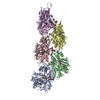
| Title | Cryo-EM structure of recombinant wildtype ACTA1 phalloidin-stabilized F-actin | ||||||||||||||||||||||||||||||||||||||||||||||||
 Components Components |
| ||||||||||||||||||||||||||||||||||||||||||||||||
 Keywords Keywords | STRUCTURAL PROTEIN / Actin / cardiomyopathy / contractility / muscle / sarcomere | ||||||||||||||||||||||||||||||||||||||||||||||||
| Function / homology |  Function and homology information Function and homology informationFormation of the dystrophin-glycoprotein complex (DGC) / Striated Muscle Contraction / Regulation of CDH1 Function / myosin binding / mesenchyme migration / striated muscle thin filament / skeletal muscle thin filament assembly / skeletal muscle fiber development / stress fiber / muscle contraction ...Formation of the dystrophin-glycoprotein complex (DGC) / Striated Muscle Contraction / Regulation of CDH1 Function / myosin binding / mesenchyme migration / striated muscle thin filament / skeletal muscle thin filament assembly / skeletal muscle fiber development / stress fiber / muscle contraction / sarcomere / filopodium / actin filament / ADP binding / structural constituent of cytoskeleton / Hydrolases; Acting on acid anhydrides; Acting on acid anhydrides to facilitate cellular and subcellular movement / lamellipodium / actin cytoskeleton / cell body / blood microparticle / hydrolase activity / positive regulation of gene expression / : / extracellular exosome / ATP binding / cytosol Similarity search - Function | ||||||||||||||||||||||||||||||||||||||||||||||||
| Biological species |  Homo sapiens (human) Homo sapiens (human) Amanita phalloides (death cap) Amanita phalloides (death cap) | ||||||||||||||||||||||||||||||||||||||||||||||||
| Method | ELECTRON MICROSCOPY / helical reconstruction / cryo EM / Resolution: 3.4 Å | ||||||||||||||||||||||||||||||||||||||||||||||||
 Authors Authors | Garg, A. / Greenberg, M.J. / Zhang, R. | ||||||||||||||||||||||||||||||||||||||||||||||||
| Funding support |  United States, United States,  France, 15items France, 15items
| ||||||||||||||||||||||||||||||||||||||||||||||||
 Citation Citation | Journal: Proc Natl Acad Sci U S A / Year: 2024 Title: Dilated cardiomyopathy-associated skeletal muscle actin (ACTA1) mutation R256H disrupts actin structure and function and causes cardiomyocyte hypocontractility. Authors: Ankit Garg / Silvia Jansen / Lina Greenberg / Rui Zhang / Kory J Lavine / Michael J Greenberg / Abstract: Skeletal muscle actin (ACTA1) mutations are a prevalent cause of skeletal myopathies consistent with ACTA1's high expression in skeletal muscle. Rare de novo mutations in ACTA1 associated with ...Skeletal muscle actin (ACTA1) mutations are a prevalent cause of skeletal myopathies consistent with ACTA1's high expression in skeletal muscle. Rare de novo mutations in ACTA1 associated with combined cardiac and skeletal myopathies have been reported, but ACTA1 represents only ~20% of the total actin pool in cardiomyocytes, making its role in cardiomyopathy controversial. Here we demonstrate how a mutation in an actin isoform expressed at low levels in cardiomyocytes can cause cardiomyopathy by focusing on a unique ACTA1 variant, R256H. We previously identified this variant in a family with dilated cardiomyopathy, who had reduced systolic function without clinical skeletal myopathy. Using a battery of multiscale biophysical tools, we show that R256H has potent effects on ACTA1 function at the molecular scale and in human cardiomyocytes. Importantly, we demonstrate that R256H acts in a dominant manner, where the incorporation of small amounts of mutant protein into thin filaments is sufficient to disrupt molecular contractility, and that this effect is dependent on the presence of troponin and tropomyosin. To understand the structural basis of this change in regulation, we resolved a structure of R256H filaments using cryoelectron microscopy, and we see alterations in actin's structure that have the potential to disrupt interactions with tropomyosin. Finally, we show that human-induced pluripotent stem cell cardiomyocytes demonstrate reduced contractility and sarcomeric organization. Taken together, we demonstrate that R256H has multiple effects on ACTA1 function that are sufficient to cause reduced contractility and establish a likely causative relationship between ACTA1 R256H and clinical cardiomyopathy. | ||||||||||||||||||||||||||||||||||||||||||||||||
| History |
|
- Structure visualization
| Structure viewer | Molecule: MolmilJmol/JSmol |
|---|
- Downloads & links
-Download
| PDBx/mmCIF format | 9duu.cif.gz | 439.4 KB | Display | PDBx/mmCIF format |
|---|---|---|---|---|
| PDB format | pdb9duu.ent.gz | 362.1 KB | Display | PDB format |
| PDBx/mmJSON format | 9duu.json.gz | Tree view | PDBx/mmJSON format | |
| Others |  Other downloads Other downloads |
-Validation report
| Arichive directory | https://data.pdbj.org/pub/pdb/validation_reports/du/9duuftp://data.pdbj.org/pub/pdb/validation_reports/du/9duu | HTTPS FTP |
|---|
-Related structure data
| Related structure data |  47179MC  9duvC M: map data used to model this data C: citing same article ( |
|---|---|
| Similar structure data |

-Links
 PDBj
PDBj


- Assembly
| Deposited unit | 
|
|---|---|
| 1 |
|
-Components
| #1: Protein | Mass: 41875.633 Da / Num. of mol.: 6 Source method: isolated from a genetically manipulated source Source: (gene. exp.) Homo sapiens (human) / Gene: ACTA1, ACTA / Production host:   Spodoptera frugiperda (fall armyworm) Spodoptera frugiperda (fall armyworm)References: UniProt: P68133, Hydrolases; Acting on acid anhydrides; Acting on acid anhydrides to facilitate cellular and subcellular movement #2: Protein/peptide | Mass: 808.899 Da / Num. of mol.: 7 / Source method: obtained synthetically / Source: (synth.) Amanita phalloides (death cap)#3: Chemical | ChemComp-ADP /   Mass: 427.201 Da / Num. of mol.: 6 / Source method: obtained synthetically / Formula: C10H15N5O10P2 / Feature type: SUBJECT OF INVESTIGATION / Comment: ADP, energy-carrying molecule*YM Mass: 427.201 Da / Num. of mol.: 6 / Source method: obtained synthetically / Formula: C10H15N5O10P2 / Feature type: SUBJECT OF INVESTIGATION / Comment: ADP, energy-carrying molecule*YM#4: Chemical | ChemComp-MG /   Mass: 24.305 Da / Num. of mol.: 6 / Source method: obtained synthetically / Formula: Mg / Feature type: SUBJECT OF INVESTIGATION Mass: 24.305 Da / Num. of mol.: 6 / Source method: obtained synthetically / Formula: Mg / Feature type: SUBJECT OF INVESTIGATIONHas ligand of interest | Y | Has protein modification | Y | |
|---|
-Experimental details
-Experiment
| Experiment | Method: ELECTRON MICROSCOPY |
|---|---|
| EM experiment | Aggregation state: FILAMENT / 3D reconstruction method: helical reconstruction |
- Sample preparation
| Component |
| ||||||||||||||||||||||||||||||
|---|---|---|---|---|---|---|---|---|---|---|---|---|---|---|---|---|---|---|---|---|---|---|---|---|---|---|---|---|---|---|---|
| Molecular weight | Experimental value: NO | ||||||||||||||||||||||||||||||
| Source (natural) |
| ||||||||||||||||||||||||||||||
| Source (recombinant) | Organism: Spodoptera frugiperda (fall armyworm) | ||||||||||||||||||||||||||||||
| Buffer solution | pH: 7 Details: 25 mM KCl, 2 mM EGTA, 60 mM MOPS (pH7), 1 mM DTT, and 4 mM MgCl2 | ||||||||||||||||||||||||||||||
| Buffer component |
| ||||||||||||||||||||||||||||||
| Specimen | Embedding applied: NO / Shadowing applied: NO / Staining applied: NO / Vitrification applied: YES | ||||||||||||||||||||||||||||||
| Specimen support | Grid material: COPPER / Grid mesh size: 400 divisions/in. / Grid type: PELCO Ultrathin Carbon with Lacey Carbon | ||||||||||||||||||||||||||||||
| Vitrification | Instrument: FEI VITROBOT MARK IV / Cryogen name: ETHANE / Humidity: 95 % / Chamber temperature: 277.15 K |
- Electron microscopy imaging
| Microscopy | Model: TFS GLACIOS |
|---|---|
| Electron gun | Electron source:  FIELD EMISSION GUN / Accelerating voltage: 200 kV / Illumination mode: FLOOD BEAM FIELD EMISSION GUN / Accelerating voltage: 200 kV / Illumination mode: FLOOD BEAM |
| Electron lens | Mode: BRIGHT FIELD / Nominal magnification: 120000 X / Nominal defocus max: 2400 nm / Nominal defocus min: 800 nm / Cs: 2.7 mm / Alignment procedure: COMA FREE |
| Specimen holder | Cryogen: NITROGEN / Specimen holder model: FEI TITAN KRIOS AUTOGRID HOLDER |
| Image recording | Average exposure time: 11.63 sec. / Electron dose: 56.7 e/Å2 / Film or detector model: FEI FALCON IV (4k x 4k) / Num. of grids imaged: 1 / Num. of real images: 2709 |
- Processing
| EM software |
| ||||||||||||||||||||||||||||||||||||||||
|---|---|---|---|---|---|---|---|---|---|---|---|---|---|---|---|---|---|---|---|---|---|---|---|---|---|---|---|---|---|---|---|---|---|---|---|---|---|---|---|---|---|
| CTF correction | Type: PHASE FLIPPING AND AMPLITUDE CORRECTION | ||||||||||||||||||||||||||||||||||||||||
| Helical symmerty | Angular rotation/subunit: -166.9 ° / Axial rise/subunit: 27.557 Å / Axial symmetry: C1 | ||||||||||||||||||||||||||||||||||||||||
| Particle selection | Details: Actin filaments were automatically picked from the micrographs using template based 'Filament tracer' in CryoSPARC | ||||||||||||||||||||||||||||||||||||||||
| 3D reconstruction | Resolution: 3.4 Å / Resolution method: FSC 0.143 CUT-OFF / Num. of particles: 1574745 / Algorithm: FOURIER SPACE / Symmetry type: HELICAL | ||||||||||||||||||||||||||||||||||||||||
| Atomic model building | Protocol: AB INITIO MODEL / Space: RECIPROCAL | ||||||||||||||||||||||||||||||||||||||||
| Atomic model building | PDB-ID: 6T1Y Accession code: 6T1Y / Source name: PDB / Type: experimental model |