[English] 日本語
Yorodumi
- PDB-9due: Crystal Structure of Human DAPK1 Catalytic Subunit Complexed with... -

+
Open data


ID or keywords:

Loading...

-
Basic information

Entry
Database: PDB / ID: 9due
TitleCrystal Structure of Human DAPK1 Catalytic Subunit Complexed with Compound SRM-07-081a
ComponentsDeath-associated protein kinase 1
KeywordsTRANSFERASE / DAPK1 / SRM-7-081a / 3-chloro-6-(4-methylpiperazin-1-yl)-4-(pyridin-4-yl)pyridazine
Function / homology
Function and homology information


cellular response to hydroperoxide / regulation of response to tumor cell / positive regulation of autophagic cell death / DAPK1-calmodulin complex / defense response to tumor cell / Caspase activation via Dependence Receptors in the absence of ligand / regulation of NMDA receptor activity / calcium/calmodulin-dependent protein kinase activity / syntaxin-1 binding / positive regulation of cysteine-type endopeptidase activity involved in apoptotic process ...cellular response to hydroperoxide / regulation of response to tumor cell / positive regulation of autophagic cell death / DAPK1-calmodulin complex / defense response to tumor cell / Caspase activation via Dependence Receptors in the absence of ligand / regulation of NMDA receptor activity / calcium/calmodulin-dependent protein kinase activity / syntaxin-1 binding / positive regulation of cysteine-type endopeptidase activity involved in apoptotic process / extrinsic apoptotic signaling pathway via death domain receptors / positive regulation of autophagy / regulation of autophagy / apoptotic signaling pathway / cellular response to type II interferon / actin cytoskeleton / regulation of apoptotic process / protein autophosphorylation / postsynaptic density / negative regulation of translation / non-specific serine/threonine protein kinase / calmodulin binding / protein kinase activity / intracellular signal transduction / positive regulation of apoptotic process / protein phosphorylation / protein serine kinase activity / protein serine/threonine kinase activity / glutamatergic synapse / GTP binding / negative regulation of apoptotic process / apoptotic process / ATP binding / identical protein binding / nucleus / plasma membrane / cytoplasm
Similarity search - Function
Death-associated protein kinase 1 / Roc domain profile. / Roc domain / Death domain profile. / DEATH domain, found in proteins involved in cell death (apoptosis). / Death domain / Death domain / Death-like domain superfamily / Ankyrin repeat / Ankyrin repeats (3 copies) ...Death-associated protein kinase 1 / Roc domain profile. / Roc domain / Death domain profile. / DEATH domain, found in proteins involved in cell death (apoptosis). / Death domain / Death domain / Death-like domain superfamily / Ankyrin repeat / Ankyrin repeats (3 copies) / Ankyrin repeat profile. / Ankyrin repeat region circular profile. / ankyrin repeats / Ankyrin repeat / Ankyrin repeat-containing domain superfamily / Serine/threonine-protein kinase, active site / Serine/Threonine protein kinases active-site signature. / Protein kinase domain / Serine/Threonine protein kinases, catalytic domain / Protein kinase, ATP binding site / Protein kinases ATP-binding region signature. / Protein kinase domain profile. / Protein kinase domain / Protein kinase-like domain superfamily / P-loop containing nucleoside triphosphate hydrolase
Similarity search - Domain/homology
: / BORIC ACID / Death-associated protein kinase 1
Similarity search - Component
Biological speciesHomo sapiens (human)
MethodX-RAY DIFFRACTION / SYNCHROTRON / MOLECULAR REPLACEMENT / Resolution: 1.65 Å
AuthorsMinasov, G. / Winsor, J. / Roy, S.M. / Watterson, D.M. / Shuvalova, L.
Funding support United States, 1items
OrganizationGrant numberCountry
National Institutes of Health/National Institute on Aging (NIH/NIA)AG066722 United States
CitationJournal: To Be Published
Title: Crystal Structure of Human DAPK1 Catalytic Subunit Complexed with Compound SRM-07-081a
Authors: Minasov, G. / Winsor, J. / Roy, S.M. / Watterson, D.M. / Shuvalova, L.
History
DepositionOct 2, 2024Deposition site: RCSB / Processing site: RCSB
Revision 1.0Oct 9, 2024Provider: repository / Type: Initial release

-
Structure visualization

Structure viewerMolecule:
MolmilJmol/JSmol

Downloads & links

-
Assembly

Deposited unit
A: Death-associated protein kinase 1
hetero molecules


Theoretical massNumber of molelcules
Total (without water)34,6268
Polymers33,7941
Non-polymers8327
Water3,639202
1


  • Idetical with deposited unit
  • defined by author&software
  • Evidence: homology, 4YPD
TypeNameSymmetry operationNumber
identity operation1_555x,y,z1
MethodPISA
Unit cell
Length a, b, c (Å)47.010, 62.684, 88.462
Angle α, β, γ (deg.)90.00, 90.00, 90.00
Int Tables number19
Space group name H-MP212121

-
Components

#1: Protein Death-associated protein kinase 1 / DAP kinase 1


Mass: 33794.367 Da / Num. of mol.: 1 / Fragment: protein kinase domain (UNP residues 2-285)
Source method: isolated from a genetically manipulated source
Source: (gene. exp.) Homo sapiens (human) / Gene: DAPK1, DAPK / Plasmid: pASK-IBA3 / Production host: Escherichia coli (E. coli)
References: UniProt: P53355, non-specific serine/threonine protein kinase
#2: Chemical ChemComp-A1BB6 / 3-chloro-6-(4-methylpiperazin-1-yl)-4-(pyridin-4-yl)pyridazine


Mass: 289.763 Da / Num. of mol.: 1 / Source method: obtained synthetically / Formula: C14H16ClN5 / Feature type: SUBJECT OF INVESTIGATION
#3: Chemical ChemComp-BO3 / BORIC ACID


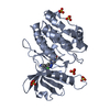
Mass: 61.833 Da / Num. of mol.: 1 / Source method: obtained synthetically / Formula: BH3O3
#4: Chemical
ChemComp-SO4 / SULFATE ION


Mass: 96.063 Da / Num. of mol.: 5 / Source method: obtained synthetically / Formula: SO4
#5: Water ChemComp-HOH / water


Mass: 18.015 Da / Num. of mol.: 202 / Source method: isolated from a natural source / Formula: H2O
Has ligand of interestY
Has protein modificationN

-
Experimental details

-
Experiment

ExperimentMethod: X-RAY DIFFRACTION / Number of used crystals: 1

-
Sample preparation

CrystalDensity Matthews: 1.93 Å3/Da / Density % sol: 36.22 %
Crystal growTemperature: 292 K / Method: vapor diffusion, hanging drop
Details: Protein: 6.6 mg/ml, 0.25M Sodium chloride, 0.1M Tris-HCl (pH 8.0); Screen: 1.8M Ammomium sulfate, 0.2M Ammonium chloride; Soak: 24h, 1mM SRM-7-081a, 10% DMSO, 1.8M Ammonium sulfate, 0.2M ...Details: Protein: 6.6 mg/ml, 0.25M Sodium chloride, 0.1M Tris-HCl (pH 8.0); Screen: 1.8M Ammomium sulfate, 0.2M Ammonium chloride; Soak: 24h, 1mM SRM-7-081a, 10% DMSO, 1.8M Ammonium sulfate, 0.2M Ammonium chloride; Cryo: 1.8M Ammonium sulfate, 25% sucrose.

-
Data collection

DiffractionMean temperature: 100 K / Serial crystal experiment: N
Diffraction sourceSource: SYNCHROTRON / Site: APS / Beamline: 21-ID-G / Wavelength: 0.97856 Å
DetectorType: MARMOSAIC 300 mm CCD / Detector: CCD / Date: Nov 14, 2019
RadiationMonochromator: C(111) / Protocol: SINGLE WAVELENGTH / Monochromatic (M) / Laue (L): M / Scattering type: x-ray
Radiation wavelengthWavelength: 0.97856 Å / Relative weight: 1
ReflectionResolution: 1.65→30 Å / Num. obs: 32407 / % possible obs: 100 % / Observed criterion σ(I): -3 / Redundancy: 7.2 % / Biso Wilson estimate: 22.3 Å2 / CC1/2: 0.998 / CC star: 0.999 / Rmerge(I) obs: 0.057 / Rpim(I) all: 0.023 / Rrim(I) all: 0.062 / Rsym value: 0.057 / Χ2: 1.004 / Net I/σ(I): 28.3
Reflection shellResolution: 1.65→1.68 Å / Redundancy: 6.3 % / Rmerge(I) obs: 0.881 / Mean I/σ(I) obs: 2.1 / Num. unique obs: 1594 / CC1/2: 0.683 / CC star: 0.901 / Rpim(I) all: 0.378 / Rrim(I) all: 0.961 / Rsym value: 0.881 / Χ2: 1.005 / % possible all: 100

-
Processing

Software
NameVersionClassification
REFMAC5.8.0425refinement
HKL-30002.3.15data reduction
HKL-30002.3.15data scaling
PHASER2.8.3phasing
RefinementMethod to determine structure: MOLECULAR REPLACEMENT / Resolution: 1.65→29.54 Å / Cor.coef. Fo:Fc: 0.972 / Cor.coef. Fo:Fc free: 0.963 / SU B: 3.575 / SU ML: 0.059 / Cross valid method: THROUGHOUT / ESU R: 0.092 / ESU R Free: 0.09 / Stereochemistry target values: MAXIMUM LIKELIHOOD / Details: HYDROGENS HAVE BEEN ADDED IN THE RIDING POSITIONS
RfactorNum. reflection% reflectionSelection details
Rfree0.18352 1577 4.9 %RANDOM
Rwork0.15375 ---
obs0.15521 30598 99.97 %-
Solvent computationIon probe radii: 0.8 Å / Shrinkage radii: 0.8 Å / VDW probe radii: 1.2 Å / Solvent model: MASK
Displacement parametersBiso mean: 26.461 Å2
Baniso -1Baniso -2Baniso -3
1--0.1 Å2-0 Å20 Å2
2--0.66 Å2-0 Å2
3----0.56 Å2
Refinement stepCycle: 1 / Resolution: 1.65→29.54 Å
ProteinNucleic acidLigandSolventTotal
Num. atoms2238 0 49 202 2489
Refine LS restraints
Refine-IDTypeDev idealDev ideal targetNumber
X-RAY DIFFRACTIONr_bond_refined_d0.0040.0122432
X-RAY DIFFRACTIONr_bond_other_d0.0010.0162314
X-RAY DIFFRACTIONr_angle_refined_deg1.5361.8523306
X-RAY DIFFRACTIONr_angle_other_deg0.5031.7795353
X-RAY DIFFRACTIONr_dihedral_angle_1_deg3.4965298
X-RAY DIFFRACTIONr_dihedral_angle_2_deg1.946513
X-RAY DIFFRACTIONr_dihedral_angle_3_deg7.61610441
X-RAY DIFFRACTIONr_dihedral_angle_4_deg
X-RAY DIFFRACTIONr_chiral_restr0.0810.2368
X-RAY DIFFRACTIONr_gen_planes_refined0.0180.022836
X-RAY DIFFRACTIONr_gen_planes_other0.0150.02545
X-RAY DIFFRACTIONr_nbd_refined
X-RAY DIFFRACTIONr_nbd_other
X-RAY DIFFRACTIONr_nbtor_refined
X-RAY DIFFRACTIONr_nbtor_other
X-RAY DIFFRACTIONr_xyhbond_nbd_refined
X-RAY DIFFRACTIONr_xyhbond_nbd_other
X-RAY DIFFRACTIONr_metal_ion_refined
X-RAY DIFFRACTIONr_metal_ion_other
X-RAY DIFFRACTIONr_symmetry_vdw_refined
X-RAY DIFFRACTIONr_symmetry_vdw_other
X-RAY DIFFRACTIONr_symmetry_hbond_refined
X-RAY DIFFRACTIONr_symmetry_hbond_other
X-RAY DIFFRACTIONr_symmetry_metal_ion_refined
X-RAY DIFFRACTIONr_symmetry_metal_ion_other
X-RAY DIFFRACTIONr_mcbond_it2.0051.8751162
X-RAY DIFFRACTIONr_mcbond_other2.0051.8741162
X-RAY DIFFRACTIONr_mcangle_it2.9543.361470
X-RAY DIFFRACTIONr_mcangle_other2.9543.3641471
X-RAY DIFFRACTIONr_scbond_it4.0282.5021270
X-RAY DIFFRACTIONr_scbond_other3.8512.4221251
X-RAY DIFFRACTIONr_scangle_it
X-RAY DIFFRACTIONr_scangle_other6.0924.1821804
X-RAY DIFFRACTIONr_long_range_B_refined7.83425.32762
X-RAY DIFFRACTIONr_long_range_B_other7.61223.612710
X-RAY DIFFRACTIONr_rigid_bond_restr
X-RAY DIFFRACTIONr_sphericity_free
X-RAY DIFFRACTIONr_sphericity_bonded
LS refinement shellResolution: 1.65→1.693 Å / Total num. of bins used: 20
RfactorNum. reflection% reflection
Rfree0.267 100 -
Rwork0.228 2246 -
obs--100 %
Refinement TLS params.

Method: refined / Refine-ID: X-RAY DIFFRACTION

IDL112)L122)L132)L222)L232)L332)S11 (Å °)S12 (Å °)S13 (Å °)S21 (Å °)S22 (Å °)S23 (Å °)S31 (Å °)S32 (Å °)S33 (Å °)T112)T122)T132)T222)T232)T332)Origin x (Å)Origin y (Å)Origin z (Å)
12.5347-1.69040.96982.7187-1.87852.4190.12530.1382-0.1915-0.1468-0.04560.2810.23210.0804-0.07970.06810.0027-0.01520.0158-0.00080.0656-2.4545-4.2635-2.5439
20.73920.3522-0.64770.6779-0.3740.58150.00360.07030.02530.02520.01950.02790.0067-0.062-0.02320.0265-0.0066-0.00370.01470.00840.01094.90528.7924-9.6502
33.5023-0.2304-0.1341.28710.35470.46990.04060.06280.13290.0247-0.01580.1160.0084-0.1436-0.02480.0151-0.00230.00020.05280.02170.02616.791111.828-10.5816
40.7585-0.13980.15710.9640.14221.25890.02450.0431-0.0179-0.03920.0339-0.0255-0.01970.0644-0.05840.03270.00060.00380.0068-0.00020.032119.263611.6354-21.5997
Refinement TLS group
IDRefine-IDRefine TLS-IDAuth asym-IDAuth seq-ID
1X-RAY DIFFRACTION1A2 - 54
2X-RAY DIFFRACTION2A55 - 149
3X-RAY DIFFRACTION3A150 - 172
4X-RAY DIFFRACTION4A173 - 277

+
About Yorodumi

-
News

-
Feb 9, 2022. New format data for meta-information of EMDB entries

New format data for meta-information of EMDB entries

  • Version 3 of the EMDB header file is now the official format.
  • The previous official version 1.9 will be removed from the archive.

Related info.:EMDB header

External links:wwPDB to switch to version 3 of the EMDB data model

-
Aug 12, 2020. Covid-19 info

Covid-19 info

URL: https://pdbjlvh1.pdbj.org/emnavi/covid19.php

New page: Covid-19 featured information page in EM Navigator.

Related info.:Covid-19 info / Mar 5, 2020. Novel coronavirus structure data

+
Mar 5, 2020. Novel coronavirus structure data

Novel coronavirus structure data

Related info.:Yorodumi Speices / Aug 12, 2020. Covid-19 info

External links:COVID-19 featured content - PDBj / Molecule of the Month (242):Coronavirus Proteases

+
Jan 31, 2019. EMDB accession codes are about to change! (news from PDBe EMDB page)

EMDB accession codes are about to change! (news from PDBe EMDB page)

  • The allocation of 4 digits for EMDB accession codes will soon come to an end. Whilst these codes will remain in use, new EMDB accession codes will include an additional digit and will expand incrementally as the available range of codes is exhausted. The current 4-digit format prefixed with “EMD-” (i.e. EMD-XXXX) will advance to a 5-digit format (i.e. EMD-XXXXX), and so on. It is currently estimated that the 4-digit codes will be depleted around Spring 2019, at which point the 5-digit format will come into force.
  • The EM Navigator/Yorodumi systems omit the EMD- prefix.

Related info.:Q: What is EMD? / ID/Accession-code notation in Yorodumi/EM Navigator

External links:EMDB Accession Codes are Changing Soon! / Contact to PDBj

+
Jul 12, 2017. Major update of PDB

Major update of PDB

  • wwPDB released updated PDB data conforming to the new PDBx/mmCIF dictionary.
  • This is a major update changing the version number from 4 to 5, and with Remediation, in which all the entries are updated.
  • In this update, many items about electron microscopy experimental information are reorganized (e.g. em_software).
  • Now, EM Navigator and Yorodumi are based on the updated data.

External links:wwPDB Remediation / Enriched Model Files Conforming to OneDep Data Standards Now Available in the PDB FTP Archive

-
Yorodumi

Thousand views of thousand structures

  • Yorodumi is a browser for structure data from EMDB, PDB, SASBDB, etc.
  • This page is also the successor to EM Navigator detail page, and also detail information page/front-end page for Omokage search.
  • The word "yorodu" (or yorozu) is an old Japanese word meaning "ten thousand". "mi" (miru) is to see.

Related info.:EMDB / PDB / SASBDB / Comparison of 3 databanks / Yorodumi Search / Aug 31, 2016. New EM Navigator & Yorodumi / Yorodumi Papers / Jmol/JSmol / Function and homology information / Changes in new EM Navigator and Yorodumi

Read more