 Movie
Movie Controller


+ Open data

- Basic information
| Entry | Database: PDB / ID: 9dti | ||||||
|---|---|---|---|---|---|---|---|
| Title | F33Y CuBMb | ||||||
 Components Components | Myoglobin | ||||||
 Keywords Keywords | OXIDOREDUCTASE / oxidase / metalloenzyme | ||||||
| Function / homology |  Function and homology information Function and homology informationOxidoreductases; Acting on other nitrogenous compounds as donors / nitrite reductase activity / sarcoplasm / Oxidoreductases; Acting on a peroxide as acceptor; Peroxidases / removal of superoxide radicals / oxygen carrier activity / peroxidase activity / oxygen binding / heme binding / extracellular exosome / metal ion binding Similarity search - Function | ||||||
| Biological species |  | ||||||
| Method |  X-RAY DIFFRACTION / SYNCHROTRON / MOLECULAR REPLACEMENT / Resolution: 1.681 Å X-RAY DIFFRACTION / SYNCHROTRON / MOLECULAR REPLACEMENT / Resolution: 1.681 Å | ||||||
 Authors Authors | Lu, Y. / Liu, Y. | ||||||
| Funding support |  United States, 1items United States, 1items
| ||||||
 Citation Citation | Journal: J.Am.Chem.Soc. / Year: 2025 Title: A Post-translational Histidine-Histidine Cross-Link Enhances Enzymatic Oxygen Reduction Activity with Greater pH Adaptability. Authors: Liu, Y. / Vilbert, A.C. / Ghosh, B. / Young, R.P. / Merkley, E.D. / Mukherjee, A. / Phan, L. / Van Stappen, C. / Baghi-Damodaran, A. / Miner, K.D. / Adkins, J. / Cort, J. / Lu, Y. | ||||||
| History |
|
- Structure visualization
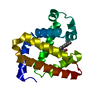
| Structure viewer | Molecule: MolmilJmol/JSmol |
|---|
- Downloads & links
-Download
| PDBx/mmCIF format | 9dti.cif.gz | 60.6 KB | Display | PDBx/mmCIF format |
|---|---|---|---|---|
| PDB format | pdb9dti.ent.gz | 38.2 KB | Display | PDB format |
| PDBx/mmJSON format | 9dti.json.gz | Tree view | PDBx/mmJSON format | |
| Others |  Other downloads Other downloads |
-Validation report
| Summary document | 9dti_validation.pdf.gz | 867.5 KB | Display | wwPDB validaton report |
|---|---|---|---|---|
| Full document | 9dti_full_validation.pdf.gz | 868.7 KB | Display | |
| Data in XML | 9dti_validation.xml.gz | 13.8 KB | Display | |
| Data in CIF | 9dti_validation.cif.gz | 19.9 KB | Display | |
| Arichive directory | https://data.pdbj.org/pub/pdb/validation_reports/dt/9dtiftp://data.pdbj.org/pub/pdb/validation_reports/dt/9dti | HTTPS FTP |
-Related structure data

-Links
 PDBj
PDBj


- Assembly
| Deposited unit | 
| ||||||||||||
|---|---|---|---|---|---|---|---|---|---|---|---|---|---|
| 1 |
| ||||||||||||
| Unit cell |
|
-Components
-Protein , 1 types, 1 molecules A
| #1: Protein | Mass: 17398.109 Da / Num. of mol.: 1 Source method: isolated from a genetically manipulated source Source: (gene. exp.)  References: UniProt: P02185, Oxidoreductases; Acting on other nitrogenous compounds as donors, Oxidoreductases; Acting on a peroxide as acceptor; Peroxidases |
|---|
-Non-polymers , 5 types, 277 molecules 


| #2: Chemical | ChemComp-HEM /  Mass: 616.487 Da / Num. of mol.: 1 / Source method: obtained synthetically / Formula: C34H32FeN4O4 / Feature type: SUBJECT OF INVESTIGATION Mass: 616.487 Da / Num. of mol.: 1 / Source method: obtained synthetically / Formula: C34H32FeN4O4 / Feature type: SUBJECT OF INVESTIGATION |
|---|---|
| #3: Chemical | ChemComp-PEG /  Mass: 106.120 Da / Num. of mol.: 1 / Source method: obtained synthetically / Formula: C4H10O3 Mass: 106.120 Da / Num. of mol.: 1 / Source method: obtained synthetically / Formula: C4H10O3 |
| #4: Chemical | ChemComp-SO4 /  Mass: 96.063 Da / Num. of mol.: 1 / Source method: obtained synthetically / Formula: SO4 Mass: 96.063 Da / Num. of mol.: 1 / Source method: obtained synthetically / Formula: SO4 |
| #5: Chemical | ChemComp-CL /  Mass: 35.453 Da / Num. of mol.: 1 / Source method: obtained synthetically / Formula: Cl Mass: 35.453 Da / Num. of mol.: 1 / Source method: obtained synthetically / Formula: Cl |
| #6: Water | ChemComp-HOH / Mass: 18.015 Da / Num. of mol.: 273 / Source method: isolated from a natural source / Formula: H2O |
-Details
| Has ligand of interest | Y |
|---|---|
| Has protein modification | N |
-Experimental details
-Experiment
| Experiment | Method: X-RAY DIFFRACTION / Number of used crystals: 1 |
|---|
- Sample preparation
| Crystal | Density Matthews: 2.45 Å3/Da / Density % sol: 49.72 % / Description: Long needles, brown color |
|---|---|
| Crystal grow | Temperature: 277 K / Method: vapor diffusion, hanging drop / pH: 8.5 Details: F33Y CuBMb (2.2 mM) in 20 mM tris(hydroxymethyl)aminomethane (tris) pH 8 (pH adjusted H2SO4), was mixed 1:1 with well buffer (0.2 M sodium acetate, 0.1 M tris hydrochloride pH 8.5, with 30% ...Details: F33Y CuBMb (2.2 mM) in 20 mM tris(hydroxymethyl)aminomethane (tris) pH 8 (pH adjusted H2SO4), was mixed 1:1 with well buffer (0.2 M sodium acetate, 0.1 M tris hydrochloride pH 8.5, with 30% polyethylene glycol 4000) using the hanging drop method, with 300 uL well buffer in the well of the crystallization tray, VAPOR DIFFUSION, HANGING DROP, temperature 277K |
-Data collection
| Diffraction | Mean temperature: 100 K / Serial crystal experiment: N |
|---|---|
| Diffraction source | Source: SYNCHROTRON / Site: ALS / Beamline: 5.0.2 / Wavelength: 1 Å |
| Detector | Type: DECTRIS PILATUS3 6M / Detector: PIXEL / Date: May 8, 2024 |
| Radiation | Monochromator: Double-crystal, Si(111) / Protocol: SINGLE WAVELENGTH / Monochromatic (M) / Laue (L): M / Scattering type: x-ray |
| Radiation wavelength | Wavelength: 1 Å / Relative weight: 1 |
| Reflection | Resolution: 1.681→38.18 Å / Num. obs: 16635 / % possible obs: 85.29 % / Redundancy: 3.1 % / Biso Wilson estimate: 17.13 Å2 / CC1/2: 0.996 / CC star: 0.999 / Rmerge(I) obs: 0.05966 / Rpim(I) all: 0.03978 / Rrim(I) all: 0.0721 / Net I/σ(I): 14 |
| Reflection shell | Resolution: 1.681→1.741 Å / Redundancy: 1.9 % / Rmerge(I) obs: 0.4 / Mean I/σ(I) obs: 1.66 / Num. unique obs: 584 / CC1/2: 0.76 / CC star: 0.929 / Rpim(I) all: 0.3582 / Rrim(I) all: 0.5398 / % possible all: 30.67 |
- Processing
| Software |
| ||||||||||||||||||||||||||||||||||||||||||||||||||||||||||||||||||||||||||||||||||||
|---|---|---|---|---|---|---|---|---|---|---|---|---|---|---|---|---|---|---|---|---|---|---|---|---|---|---|---|---|---|---|---|---|---|---|---|---|---|---|---|---|---|---|---|---|---|---|---|---|---|---|---|---|---|---|---|---|---|---|---|---|---|---|---|---|---|---|---|---|---|---|---|---|---|---|---|---|---|---|---|---|---|---|---|---|---|
| Refinement | Method to determine structure: MOLECULAR REPLACEMENT / Resolution: 1.681→38.18 Å / Cross valid method: FREE R-VALUE / σ(F): 1.34 Stereochemistry target values: GeoStd + Monomer Library + CDL v1.2
| ||||||||||||||||||||||||||||||||||||||||||||||||||||||||||||||||||||||||||||||||||||
| Solvent computation | Solvent model: FLAT BULK SOLVENT MODEL | ||||||||||||||||||||||||||||||||||||||||||||||||||||||||||||||||||||||||||||||||||||
| Displacement parameters | Biso mean: 21.33 Å2 | ||||||||||||||||||||||||||||||||||||||||||||||||||||||||||||||||||||||||||||||||||||
| Refinement step | Cycle: LAST / Resolution: 1.681→38.18 Å
| ||||||||||||||||||||||||||||||||||||||||||||||||||||||||||||||||||||||||||||||||||||
| Refine LS restraints |
| ||||||||||||||||||||||||||||||||||||||||||||||||||||||||||||||||||||||||||||||||||||
| LS refinement shell |
|
