 Movie
Movie Controller

[English] 日本語
 Yorodumi
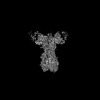
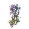
- PDB-9ds1: Crystal structure of 241_2F04 Fab in complex with H1 HA from A/Ca... -
+ Open data

- Basic information
| Entry | Database: PDB / ID: 9ds1 | ||||||
|---|---|---|---|---|---|---|---|
| Title | Crystal structure of 241_2F04 Fab in complex with H1 HA from A/California/04/2009(H1N1) | ||||||
 Components Components |
| ||||||
 Keywords Keywords | VIRAL PROTEIN/IMMUNE SYSTEM / H1N1 / Antibody / Hemagglutinin / VIRAL PROTEIN-IMMUNE SYSTEM complex / VIRAL PROTEIN | ||||||
| Function / homology |  Function and homology information Function and homology informationviral budding from plasma membrane / clathrin-dependent endocytosis of virus by host cell / host cell surface receptor binding / fusion of virus membrane with host plasma membrane / fusion of virus membrane with host endosome membrane / viral envelope / virion attachment to host cell / host cell plasma membrane / virion membrane / membrane Similarity search - Function | ||||||
| Biological species |   Influenza A virus Influenza A virus Homo sapiens (human) Homo sapiens (human) | ||||||
| Method |  X-RAY DIFFRACTION / SYNCHROTRON / MOLECULAR REPLACEMENT / Resolution: 2.4 Å X-RAY DIFFRACTION / SYNCHROTRON / MOLECULAR REPLACEMENT / Resolution: 2.4 Å | ||||||
 Authors Authors | Lin, T.H. / Wilson, I.A. | ||||||
| Funding support |  United States, 1items United States, 1items
| ||||||
 Citation Citation | Journal: Nat Commun / Year: 2025 Title: Structurally convergent antibodies derived from different vaccine strategies target the influenza virus HA anchor epitope with a subset of V3 and V3 genes. Authors: Ting-Hui Lin / Chang-Chun David Lee / Monica L Fernández-Quintero / James A Ferguson / Julianna Han / Xueyong Zhu / Wenli Yu / Jenna J Guthmiller / Florian Krammer / Patrick C Wilson / ...Authors: Ting-Hui Lin / Chang-Chun David Lee / Monica L Fernández-Quintero / James A Ferguson / Julianna Han / Xueyong Zhu / Wenli Yu / Jenna J Guthmiller / Florian Krammer / Patrick C Wilson / Andrew B Ward / Ian A Wilson /  Abstract: H1N1 influenza viruses are responsible for both seasonal and pandemic influenza. The continual antigenic shift and drift of these viruses highlight the urgent need for a universal influenza vaccine ...H1N1 influenza viruses are responsible for both seasonal and pandemic influenza. The continual antigenic shift and drift of these viruses highlight the urgent need for a universal influenza vaccine to elicit broadly neutralizing antibodies (bnAbs). Identification and characterization of bnAbs elicited in natural infection and immunization to influenza virus hemagglutinin (HA) can provide insights for development of a universal influenza vaccine. Here, we structurally and biophysically characterize four antibodies that bind to a conserved region on the HA membrane-proximal region known as the anchor epitope. Despite some diversity in their V and V genes, the antibodies interact with the HA through germline-encoded residues in HCDR2 and LCDR3. Somatic mutations on HCDR3 also contribute hydrophobic interactions with the conserved HA epitope. This convergent binding mode provides extensive neutralization breadth against H1N1 viruses and suggests possible countermeasures against H1N1 viruses. | ||||||
| History |
|
- Structure visualization
| Structure viewer | Molecule: MolmilJmol/JSmol |
|---|
- Downloads & links
-Download
| PDBx/mmCIF format | 9ds1.cif.gz | 202.6 KB | Display | PDBx/mmCIF format |
|---|---|---|---|---|
| PDB format | pdb9ds1.ent.gz | 157.4 KB | Display | PDB format |
| PDBx/mmJSON format | 9ds1.json.gz | Tree view | PDBx/mmJSON format | |
| Others |  Other downloads Other downloads |
-Validation report
| Arichive directory | https://data.pdbj.org/pub/pdb/validation_reports/ds/9ds1ftp://data.pdbj.org/pub/pdb/validation_reports/ds/9ds1 | HTTPS FTP |
|---|
-Related structure data


-Links
 PDBj
PDBj


- Assembly
| Deposited unit | 
| |||||||||
|---|---|---|---|---|---|---|---|---|---|---|
| 1 | 
| |||||||||
| Unit cell |
| |||||||||
| Components on special symmetry positions |
|
-Components
-Hemagglutinin ... , 2 types, 2 molecules AB
| #1: Protein | Mass: 36213.812 Da / Num. of mol.: 1 Source method: isolated from a genetically manipulated source Source: (gene. exp.) Influenza A virus / Gene: HA / Production host:  Trichoplusia ni (cabbage looper) / References: UniProt: A0A5B9ZSV0 Trichoplusia ni (cabbage looper) / References: UniProt: A0A5B9ZSV0 |
|---|---|
| #2: Protein | Mass: 19992.076 Da / Num. of mol.: 1 Source method: isolated from a genetically manipulated source Source: (gene. exp.) Influenza A virus / Gene: HA / Production host: Trichoplusia ni (cabbage looper) / References: UniProt: A0A6J3XB93 |
-Antibody , 2 types, 2 molecules HL
| #3: Antibody | Mass: 24169.281 Da / Num. of mol.: 1 Source method: isolated from a genetically manipulated source Source: (gene. exp.) Homo sapiens (human) / Production host:   Cricetulus griseus (Chinese hamster) Cricetulus griseus (Chinese hamster) |
|---|---|
| #4: Antibody | Mass: 23493.066 Da / Num. of mol.: 1 Source method: isolated from a genetically manipulated source Source: (gene. exp.) Homo sapiens (human) / Production host: Cricetulus griseus (Chinese hamster) |
-Sugars , 2 types, 2 molecules 
| #5: Polysaccharide | beta-D-mannopyranose-(1-4)-2-acetamido-2-deoxy-beta-D-glucopyranose-(1-4)-2-acetamido-2-deoxy-beta- ...beta-D-mannopyranose-(1-4)-2-acetamido-2-deoxy-beta-D-glucopyranose-(1-4)-2-acetamido-2-deoxy-beta-D-glucopyranose Source method: isolated from a genetically manipulated source |
|---|---|
| #6: Sugar | ChemComp-NAG /  Type: D-saccharide, beta linking / Mass: 221.208 Da / Num. of mol.: 1 / Source method: obtained synthetically / Formula: C8H15NO6 / Feature type: SUBJECT OF INVESTIGATION Type: D-saccharide, beta linking / Mass: 221.208 Da / Num. of mol.: 1 / Source method: obtained synthetically / Formula: C8H15NO6 / Feature type: SUBJECT OF INVESTIGATION |
-Non-polymers , 1 types, 129 molecules 
| #7: Water | ChemComp-HOH / Mass: 18.015 Da / Num. of mol.: 129 / Source method: isolated from a natural source / Formula: H2O |
|---|
-Details
| Has ligand of interest | Y |
|---|---|
| Has protein modification | Y |
-Experimental details
-Experiment
| Experiment | Method: X-RAY DIFFRACTION / Number of used crystals: 1 |
|---|
- Sample preparation
| Crystal | Density Matthews: 3.45 Å3/Da / Density % sol: 64.3 % |
|---|---|
| Crystal grow | Temperature: 293 K / Method: vapor diffusion, hanging drop / Details: 0.2 M sodium acetate, pH 6.4, 16% PEG3350 |
-Data collection
| Diffraction | Mean temperature: 100 K / Serial crystal experiment: N |
|---|---|
| Diffraction source | Source: SYNCHROTRON / Site: SSRL / Beamline: BL12-1 / Wavelength: 0.97934 Å |
| Detector | Type: DECTRIS EIGER X 16M / Detector: PIXEL / Date: Nov 16, 2021 |
| Radiation | Protocol: SINGLE WAVELENGTH / Monochromatic (M) / Laue (L): M / Scattering type: x-ray |
| Radiation wavelength | Wavelength: 0.97934 Å / Relative weight: 1 |
| Reflection | Resolution: 2.4→39.43 Å / Num. obs: 54268 / % possible obs: 96 % / Redundancy: 19 % / CC1/2: 1 / Rmerge(I) obs: 0.18 / Rpim(I) all: 0.04 / Net I/σ(I): 15 |
| Reflection shell | Resolution: 2.4→2.44 Å / Rmerge(I) obs: 1.1 / Num. unique obs: 54123 / CC1/2: 0.9 / Rpim(I) all: 0.28 |
- Processing
| Software |
| |||||||||||||||||||||||||||||||||||||||||||||||||||||||||||||||||||||||||||||||||||||||||||||||||||||||||||||||||||||||||||||||||||||||||||||||||||
|---|---|---|---|---|---|---|---|---|---|---|---|---|---|---|---|---|---|---|---|---|---|---|---|---|---|---|---|---|---|---|---|---|---|---|---|---|---|---|---|---|---|---|---|---|---|---|---|---|---|---|---|---|---|---|---|---|---|---|---|---|---|---|---|---|---|---|---|---|---|---|---|---|---|---|---|---|---|---|---|---|---|---|---|---|---|---|---|---|---|---|---|---|---|---|---|---|---|---|---|---|---|---|---|---|---|---|---|---|---|---|---|---|---|---|---|---|---|---|---|---|---|---|---|---|---|---|---|---|---|---|---|---|---|---|---|---|---|---|---|---|---|---|---|---|---|---|---|---|
| Refinement | Method to determine structure: MOLECULAR REPLACEMENT / Resolution: 2.4→39.43 Å / SU ML: 0.36 / Cross valid method: FREE R-VALUE / σ(F): 1.36 / Phase error: 31.37 / Stereochemistry target values: ML
| |||||||||||||||||||||||||||||||||||||||||||||||||||||||||||||||||||||||||||||||||||||||||||||||||||||||||||||||||||||||||||||||||||||||||||||||||||
| Solvent computation | Shrinkage radii: 0.9 Å / VDW probe radii: 1.1 Å / Solvent model: FLAT BULK SOLVENT MODEL | |||||||||||||||||||||||||||||||||||||||||||||||||||||||||||||||||||||||||||||||||||||||||||||||||||||||||||||||||||||||||||||||||||||||||||||||||||
| Refinement step | Cycle: LAST / Resolution: 2.4→39.43 Å
| |||||||||||||||||||||||||||||||||||||||||||||||||||||||||||||||||||||||||||||||||||||||||||||||||||||||||||||||||||||||||||||||||||||||||||||||||||
| Refine LS restraints |
| |||||||||||||||||||||||||||||||||||||||||||||||||||||||||||||||||||||||||||||||||||||||||||||||||||||||||||||||||||||||||||||||||||||||||||||||||||
| LS refinement shell |
|
