 Movie
Movie Controller

[English] 日本語
 Yorodumi
- PDB-9dr4: Crystal structure of bifunctional GlmU from Staphylococcus aureus... -
+ Open data

- Basic information
| Entry | Database: PDB / ID: 9dr4 | ||||||
|---|---|---|---|---|---|---|---|
| Title | Crystal structure of bifunctional GlmU from Staphylococcus aureus NCTC 8325 complexed with UTP, CoA and Glc 1-P | ||||||
 Components Components | Bifunctional protein GlmU | ||||||
 Keywords Keywords | TRANSFERASE / uridyltransferase / acetyltransferase | ||||||
| Function / homology |  Function and homology information Function and homology informationglucosamine-1-phosphate N-acetyltransferase / glucosamine-1-phosphate N-acetyltransferase activity / UDP-N-acetylglucosamine diphosphorylase / UDP-N-acetylglucosamine diphosphorylase activity / UDP-N-acetylglucosamine biosynthetic process / lipid A biosynthetic process / peptidoglycan biosynthetic process / cell wall organization / cell morphogenesis / regulation of cell shape ...glucosamine-1-phosphate N-acetyltransferase / glucosamine-1-phosphate N-acetyltransferase activity / UDP-N-acetylglucosamine diphosphorylase / UDP-N-acetylglucosamine diphosphorylase activity / UDP-N-acetylglucosamine biosynthetic process / lipid A biosynthetic process / peptidoglycan biosynthetic process / cell wall organization / cell morphogenesis / regulation of cell shape / magnesium ion binding / membrane / cytosol Similarity search - Function | ||||||
| Biological species |  Staphylococcus aureus subsp. aureus NCTC 8325 (bacteria) Staphylococcus aureus subsp. aureus NCTC 8325 (bacteria) | ||||||
| Method |  X-RAY DIFFRACTION / SYNCHROTRON / MOLECULAR REPLACEMENT / Resolution: 1.85 Å X-RAY DIFFRACTION / SYNCHROTRON / MOLECULAR REPLACEMENT / Resolution: 1.85 Å | ||||||
 Authors Authors | Pederick, J.L. / Bruning, J.B. | ||||||
| Funding support | 1items
| ||||||
 Citation Citation | Journal: Protein Sci. / Year: 2025 Title: Functional and structural characterization of Staphylococcus aureus N-acetylglucosamine 1-phosphate uridyltransferase (GlmU) reveals a redox-sensitive acetyltransferase activity. Authors: Pederick, J.L. / Kumar, A. / Pukala, T.L. / Bruning, J.B. | ||||||
| History |
|
- Structure visualization
| Structure viewer | Molecule: MolmilJmol/JSmol |
|---|
- Downloads & links
-Download
| PDBx/mmCIF format | 9dr4.cif.gz | 111.3 KB | Display | PDBx/mmCIF format |
|---|---|---|---|---|
| PDB format | pdb9dr4.ent.gz | Display | PDB format | |
| PDBx/mmJSON format | 9dr4.json.gz | Tree view | PDBx/mmJSON format | |
| Others |  Other downloads Other downloads |
-Validation report
| Arichive directory | https://data.pdbj.org/pub/pdb/validation_reports/dr/9dr4ftp://data.pdbj.org/pub/pdb/validation_reports/dr/9dr4 | HTTPS FTP |
|---|
-Related structure data

-Links
 PDBj
PDBj


- Assembly
| Deposited unit | 
| |||||||||||||||||||||||||||
|---|---|---|---|---|---|---|---|---|---|---|---|---|---|---|---|---|---|---|---|---|---|---|---|---|---|---|---|---|
| 1 | 
| |||||||||||||||||||||||||||
| Unit cell |
| |||||||||||||||||||||||||||
| Components on special symmetry positions |
|

-Components
-Protein / Sugars , 2 types, 2 molecules A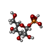
| #1: Protein | Mass: 49722.773 Da / Num. of mol.: 1 Source method: isolated from a genetically manipulated source Source: (gene. exp.) Staphylococcus aureus subsp. aureus NCTC 8325 (bacteria)Gene: glmU, SAOUHSC_00471 / Production host: References: UniProt: Q2G0S3, UDP-N-acetylglucosamine diphosphorylase, glucosamine-1-phosphate N-acetyltransferase |
|---|---|
| #4: Sugar | ChemComp-G1P /  Type: D-saccharide / Mass: 260.136 Da / Num. of mol.: 1 / Source method: obtained synthetically / Formula: C6H13O9P / Feature type: SUBJECT OF INVESTIGATION Type: D-saccharide / Mass: 260.136 Da / Num. of mol.: 1 / Source method: obtained synthetically / Formula: C6H13O9P / Feature type: SUBJECT OF INVESTIGATION |
-Non-polymers , 4 types, 265 molecules 


| #2: Chemical | ChemComp-COA /  Mass: 767.534 Da / Num. of mol.: 1 / Source method: obtained synthetically / Formula: C21H36N7O16P3S / Feature type: SUBJECT OF INVESTIGATION Mass: 767.534 Da / Num. of mol.: 1 / Source method: obtained synthetically / Formula: C21H36N7O16P3S / Feature type: SUBJECT OF INVESTIGATION |
|---|---|
| #3: Chemical | ChemComp-MG /  Mass: 24.305 Da / Num. of mol.: 1 / Source method: obtained synthetically / Formula: Mg Mass: 24.305 Da / Num. of mol.: 1 / Source method: obtained synthetically / Formula: Mg |
| #5: Chemical | ChemComp-UTP /  Mass: 484.141 Da / Num. of mol.: 1 / Source method: isolated from a natural source / Formula: C9H15N2O15P3 / Feature type: SUBJECT OF INVESTIGATION / Comment: UTP*YM Mass: 484.141 Da / Num. of mol.: 1 / Source method: isolated from a natural source / Formula: C9H15N2O15P3 / Feature type: SUBJECT OF INVESTIGATION / Comment: UTP*YM |
| #6: Water | ChemComp-HOH / Mass: 18.015 Da / Num. of mol.: 262 / Source method: isolated from a natural source / Formula: H2O |
-Details
| Has ligand of interest | Y |
|---|---|
| Has protein modification | N |
-Experimental details
-Experiment
| Experiment | Method: X-RAY DIFFRACTION / Number of used crystals: 1 |
|---|
- Sample preparation
| Crystal | Density Matthews: 2.29 Å3/Da / Density % sol: 46.22 % / Description: Trapezoid |
|---|---|
| Crystal grow | Temperature: 289.15 K / Method: vapor diffusion, hanging drop Details: 0.2M lithium citrate tribasic tetrahydrate, 15 - 20% PEG3350 |
-Data collection
| Diffraction | Mean temperature: 100 K / Serial crystal experiment: N |
|---|---|
| Diffraction source | Source: SYNCHROTRON / Site: Australian Synchrotron  / Beamline: MX2 / Wavelength: 0.9537 Å / Beamline: MX2 / Wavelength: 0.9537 Å |
| Detector | Type: DECTRIS EIGER X 16M / Detector: PIXEL / Date: Oct 27, 2019 |
| Radiation | Protocol: SINGLE WAVELENGTH / Monochromatic (M) / Laue (L): M / Scattering type: x-ray |
| Radiation wavelength | Wavelength: 0.9537 Å / Relative weight: 1 |
| Reflection | Resolution: 1.85→39.21 Å / Num. obs: 39284 / % possible obs: 100 % / Redundancy: 20.3 % / CC1/2: 0.998 / Rmerge(I) obs: 0.239 / Rpim(I) all: 0.054 / Rrim(I) all: 0.245 / Χ2: 0.99 / Net I/σ(I): 12.1 / Num. measured all: 796494 |
| Reflection shell | Resolution: 1.85→1.89 Å / % possible obs: 100 % / Redundancy: 20.4 % / Rmerge(I) obs: 3.603 / Num. measured all: 49237 / Num. unique obs: 2410 / CC1/2: 0.526 / Rpim(I) all: 0.814 / Rrim(I) all: 3.695 / Χ2: 0.95 / Net I/σ(I) obs: 1.2 |
- Processing
| Software |
| |||||||||||||||||||||||||||||||||||||||||||||||||||||||||||||||||||||||||||||||||||||||||||||||||||||||||
|---|---|---|---|---|---|---|---|---|---|---|---|---|---|---|---|---|---|---|---|---|---|---|---|---|---|---|---|---|---|---|---|---|---|---|---|---|---|---|---|---|---|---|---|---|---|---|---|---|---|---|---|---|---|---|---|---|---|---|---|---|---|---|---|---|---|---|---|---|---|---|---|---|---|---|---|---|---|---|---|---|---|---|---|---|---|---|---|---|---|---|---|---|---|---|---|---|---|---|---|---|---|---|---|---|---|---|
| Refinement | Method to determine structure: MOLECULAR REPLACEMENT / Resolution: 1.85→39.21 Å / SU ML: 0.2 / Cross valid method: FREE R-VALUE / σ(F): 1.34 / Phase error: 22.51 / Stereochemistry target values: ML
| |||||||||||||||||||||||||||||||||||||||||||||||||||||||||||||||||||||||||||||||||||||||||||||||||||||||||
| Solvent computation | Shrinkage radii: 0.9 Å / VDW probe radii: 1.1 Å / Solvent model: FLAT BULK SOLVENT MODEL | |||||||||||||||||||||||||||||||||||||||||||||||||||||||||||||||||||||||||||||||||||||||||||||||||||||||||
| Refinement step | Cycle: LAST / Resolution: 1.85→39.21 Å
| |||||||||||||||||||||||||||||||||||||||||||||||||||||||||||||||||||||||||||||||||||||||||||||||||||||||||
| Refine LS restraints |
| |||||||||||||||||||||||||||||||||||||||||||||||||||||||||||||||||||||||||||||||||||||||||||||||||||||||||
| LS refinement shell |
|
