[English] 日本語
Yorodumi
- PDB-9dqn: Nan Regulatory Protein (NanR) - DNA complex from Streptococcus pn... -

+
Open data


ID or keywords:

Loading...

-
Basic information

Entry
Database: PDB / ID: 9dqn
TitleNan Regulatory Protein (NanR) - DNA complex from Streptococcus pneumoniae
Components
  • (DNA (5'-D(P*TP*CP*TP*GP*AP*AP*AP*GP*TP*AP*CP*TP*TP*TP*TP*AP*GP*A)- ...) x 2
  • Phosphosugar-binding transcriptional regulator
KeywordsTRANSCRIPTION/DNA / Regulator / protein-DNA complex / DNA-Binding / Ligand-bound / Isomerase-Domain / TRANSCRIPTION / TRANSCRIPTION-DNA complex
Function / homology
Function and homology information


carbohydrate derivative metabolic process / carbohydrate derivative binding / DNA-binding transcription factor activity / DNA binding
Similarity search - Function
Helix-turn-helix protein RpiR / : / Helix-turn-helix domain, rpiR family / RpiR-type HTH domain profile. / RpiR-like, SIS domain / SIS domain / SIS domain / SIS domain profile. / SIS domain superfamily / Homeobox-like domain superfamily / Winged helix-like DNA-binding domain superfamily
Similarity search - Domain/homology
DNA / DNA (> 10) / Phosphosugar-binding transcriptional regulator
Similarity search - Component
Biological speciesStreptococcus pneumoniae (bacteria)
synthetic construct (others)
MethodX-RAY DIFFRACTION / SYNCHROTRON / MOLECULAR REPLACEMENT / Resolution: 2.99 Å
AuthorsWood, D.M. / Dobson, R.C.J. / Horne, C.R.
Funding support New Zealand, 2items
OrganizationGrant numberCountry
Royal Society of New ZealandUOC1506 New Zealand
Ministry of Business, Innovation and Employment (New Zealand)UOCX1706 New Zealand
CitationJournal: To Be Published
Title: Nan Regulatory Protein (NanR) - DNA complex from Streptococcus pneumoniae (CASP target)
Authors: Wood, D.M. / Dobson, R.C.J. / Horne, C.R.
History
DepositionSep 24, 2024Deposition site: RCSB / Processing site: RCSB
Revision 1.0Aug 20, 2025Provider: repository / Type: Initial release

-
Structure visualization

Structure viewerMolecule:
MolmilJmol/JSmol

Downloads & links

-
Assembly

Deposited unit
A: Phosphosugar-binding transcriptional regulator
C: Phosphosugar-binding transcriptional regulator
D: Phosphosugar-binding transcriptional regulator
B: Phosphosugar-binding transcriptional regulator
H: DNA (5'-D(P*TP*CP*TP*GP*AP*AP*AP*GP*TP*AP*CP*TP*TP*TP*TP*AP*GP*A)-3')
I: DNA (5'-D(P*TP*CP*TP*GP*AP*AP*AP*GP*TP*AP*CP*TP*TP*TP*TP*AP*GP*A)-3')
J: DNA (5'-D(P*TP*CP*TP*GP*AP*AP*AP*GP*TP*AP*CP*TP*TP*TP*TP*AP*GP*A)-3')
K: DNA (5'-D(P*TP*CP*TP*GP*AP*AP*AP*GP*TP*AP*CP*TP*TP*TP*TP*AP*GP*A)-3')
hetero molecules


Theoretical massNumber of molelcules
Total (without water)152,89312
Polymers152,6608
Non-polymers2334
Water181
1
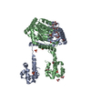
A: Phosphosugar-binding transcriptional regulator
B: Phosphosugar-binding transcriptional regulator
H: DNA (5'-D(P*TP*CP*TP*GP*AP*AP*AP*GP*TP*AP*CP*TP*TP*TP*TP*AP*GP*A)-3')
I: DNA (5'-D(P*TP*CP*TP*GP*AP*AP*AP*GP*TP*AP*CP*TP*TP*TP*TP*AP*GP*A)-3')
hetero molecules


  • defined by author
  • Evidence: SAXS, SAXS of the protein-DNA molecule suggests that homodimeric protein binds DNA as a dimer at physiological conditions, assay for oligomerization, Analytical ultracentrifuge experiments ...Evidence: SAXS, SAXS of the protein-DNA molecule suggests that homodimeric protein binds DNA as a dimer at physiological conditions, assay for oligomerization, Analytical ultracentrifuge experiments suggest that this homodimeric protein binds DNA as a dimer at physiological conditions, assay for oligomerization, The tetramer protein that forms the asymmetric unit in this crystal is the exact same tetramer conformation as a mutant protein (SpNanR Arg148) we first crystallized with DNA, and believe that it is a crystal contact, assay for oligomerization, PDBePISA suggests that the contacts formed between the two protein dimers are not inherently solution based, and the buried surface area does not corrospond to a well-define protein-protein interface
  • 76.6 kDa, 4 polymers
  • Search similar-shape structures of this assembly by Omokage search (details)
Theoretical massNumber of molelcules
Total (without water)76,5638
Polymers76,3304
Non-polymers2334
Water0
TypeNameSymmetry operationNumber
identity operation1_555x,y,z1
2
C: Phosphosugar-binding transcriptional regulator
D: Phosphosugar-binding transcriptional regulator
J: DNA (5'-D(P*TP*CP*TP*GP*AP*AP*AP*GP*TP*AP*CP*TP*TP*TP*TP*AP*GP*A)-3')
K: DNA (5'-D(P*TP*CP*TP*GP*AP*AP*AP*GP*TP*AP*CP*TP*TP*TP*TP*AP*GP*A)-3')


Theoretical massNumber of molelcules
Total (without water)76,3304
Polymers76,3304
Non-polymers00
Water181
TypeNameSymmetry operationNumber
identity operation1_555x,y,z1
Unit cell
Length a, b, c (Å)57.893, 159.393, 171.751
Angle α, β, γ (deg.)90.000, 90.000, 90.000
Int Tables number18
Space group name H-MP22121
Space group name HallP22ab(z,x,y)
Symmetry operation#1: x,y,z
#2: x,-y,-z
#3: -x,y+1/2,-z+1/2
#4: -x,-y+1/2,z+1/2

-
Components

-
Protein , 1 types, 4 molecules ACDB

#1: Protein
Phosphosugar-binding transcriptional regulator / Nan regulatory protein


Mass: 32650.963 Da / Num. of mol.: 4
Source method: isolated from a genetically manipulated source
Source: (gene. exp.) Streptococcus pneumoniae (bacteria)
Gene: ybbH, ybbH_1, SAMEA2467332_00633, SAMEA3176123_01518, SAMEA3176126_00944, SAMEA3176127_00886
Production host: Escherichia coli BL21(DE3) (bacteria) / References: UniProt: A0A4M6CQT5

-
DNA (5'-D(P*TP*CP*TP*GP*AP*AP*AP*GP*TP*AP*CP*TP*TP*TP*TP*AP*GP*A)- ... , 2 types, 4 molecules HJIK

#2: DNA chain DNA (5'-D(P*TP*CP*TP*GP*AP*AP*AP*GP*TP*AP*CP*TP*TP*TP*TP*AP*GP*A)-3')


Mass: 5529.614 Da / Num. of mol.: 2 / Source method: obtained synthetically / Source: (synth.) synthetic construct (others)
#3: DNA chain DNA (5'-D(P*TP*CP*TP*GP*AP*AP*AP*GP*TP*AP*CP*TP*TP*TP*TP*AP*GP*A)-3')


Mass: 5498.604 Da / Num. of mol.: 2 / Source method: obtained synthetically / Source: (synth.) synthetic construct (others)

-
Non-polymers , 3 types, 5 molecules

#4: Chemical ChemComp-GOL / GLYCEROL / GLYCERIN / PROPANE-1,2,3-TRIOL


Mass: 92.094 Da / Num. of mol.: 2 / Source method: obtained synthetically / Formula: C3H8O3
#5: Chemical ChemComp-MG / MAGNESIUM ION


Mass: 24.305 Da / Num. of mol.: 2 / Source method: obtained synthetically / Formula: Mg
#6: Water ChemComp-HOH / water


Mass: 18.015 Da / Num. of mol.: 1 / Source method: isolated from a natural source / Formula: H2O

-
Details

Has ligand of interestN
Has protein modificationN

-
Experimental details

-
Experiment

ExperimentMethod: X-RAY DIFFRACTION / Number of used crystals: 1

-
Sample preparation

CrystalDensity Matthews: 2.6 Å3/Da / Density % sol: 52.61 %
Crystal growTemperature: 293.15 K / Method: vapor diffusion, sitting drop
Details: SG1 E3 condition: 0.2 M Magnesium chloride hexahydrate, 0.1 M Sodium HEPES 7.5, 30 % v/v PEG 400

-
Data collection

DiffractionMean temperature: 100 K / Serial crystal experiment: N
Diffraction sourceSource: SYNCHROTRON / Site: Australian Synchrotron / Beamline: MX2 / Wavelength: 0.953739 Å
DetectorType: DECTRIS EIGER X 16M / Detector: PIXEL / Date: Nov 9, 2021
RadiationProtocol: SINGLE WAVELENGTH / Monochromatic (M) / Laue (L): M / Scattering type: x-ray
Radiation wavelengthWavelength: 0.953739 Å / Relative weight: 1
ReflectionResolution: 2.99→46 Å / Num. obs: 30263 / % possible obs: 92 % / Redundancy: 4.9 % / Biso Wilson estimate: 86.11 Å2 / CC1/2: 0.998 / Net I/σ(I): 7.9
Reflection shellResolution: 2.99→3.13 Å / Redundancy: 5.1 % / Mean I/σ(I) obs: 0.8 / Num. unique obs: 4821 / CC1/2: 0.25 / % possible all: 99.8

-
Processing

Software
NameVersionClassification
PHENIX1.21.1_5286refinement
XDSdata reduction
XDSdata scaling
PHASERphasing
RefinementMethod to determine structure: MOLECULAR REPLACEMENT / Resolution: 2.99→46 Å / SU ML: 0.4758 / Cross valid method: FREE R-VALUE / σ(F): 1.33 / Phase error: 30.0399
Stereochemistry target values: GeoStd + Monomer Library + CDL v1.2
RfactorNum. reflection% reflection
Rfree0.2598 1522 5.03 %
Rwork0.2121 28741 -
obs0.2145 30263 91.51 %
Solvent computationShrinkage radii: 0.9 Å / VDW probe radii: 1.1 Å / Solvent model: FLAT BULK SOLVENT MODEL
Displacement parametersBiso mean: 85.16 Å2
Refinement stepCycle: LAST / Resolution: 2.99→46 Å
ProteinNucleic acidLigandSolventTotal
Num. atoms8685 1476 14 1 10176
Refine LS restraints
Refine-IDTypeDev idealNumber
X-RAY DIFFRACTIONf_bond_d0.003410507
X-RAY DIFFRACTIONf_angle_d0.562414491
X-RAY DIFFRACTIONf_chiral_restr0.03691625
X-RAY DIFFRACTIONf_plane_restr0.00441615
X-RAY DIFFRACTIONf_dihedral_angle_d21.66844102
LS refinement shell
Resolution (Å)Rfactor RfreeNum. reflection RfreeRfactor RworkNum. reflection RworkRefine-ID% reflection obs (%)
2.99-3.090.42821430.3962808X-RAY DIFFRACTION99.39
3.09-3.20.38641420.30452785X-RAY DIFFRACTION99.29
3.2-3.320.30071650.27972757X-RAY DIFFRACTION99.02
3.32-3.440.36441060.27522145X-RAY DIFFRACTION94.66
3.49-3.660.26181260.23222468X-RAY DIFFRACTION96
3.66-3.890.23291010.21182050X-RAY DIFFRACTION72.23
3.89-4.190.2391240.18412212X-RAY DIFFRACTION78.23
4.19-4.610.23441600.18872813X-RAY DIFFRACTION99.53
4.61-5.280.23841410.18362878X-RAY DIFFRACTION99.31
5.28-6.640.251400.22522857X-RAY DIFFRACTION98.1
6.64-45.960.2391740.1772968X-RAY DIFFRACTION97.58

+
About Yorodumi

-
News

-
Feb 9, 2022. New format data for meta-information of EMDB entries

New format data for meta-information of EMDB entries

  • Version 3 of the EMDB header file is now the official format.
  • The previous official version 1.9 will be removed from the archive.

Related info.:EMDB header

External links:wwPDB to switch to version 3 of the EMDB data model

-
Aug 12, 2020. Covid-19 info

Covid-19 info

URL: https://pdbj.org/emnavi/covid19.php

New page: Covid-19 featured information page in EM Navigator.

Related info.:Covid-19 info / Mar 5, 2020. Novel coronavirus structure data

+
Mar 5, 2020. Novel coronavirus structure data

Novel coronavirus structure data

Related info.:Yorodumi Speices / Aug 12, 2020. Covid-19 info

External links:COVID-19 featured content - PDBj / Molecule of the Month (242):Coronavirus Proteases

+
Jan 31, 2019. EMDB accession codes are about to change! (news from PDBe EMDB page)

EMDB accession codes are about to change! (news from PDBe EMDB page)

  • The allocation of 4 digits for EMDB accession codes will soon come to an end. Whilst these codes will remain in use, new EMDB accession codes will include an additional digit and will expand incrementally as the available range of codes is exhausted. The current 4-digit format prefixed with “EMD-” (i.e. EMD-XXXX) will advance to a 5-digit format (i.e. EMD-XXXXX), and so on. It is currently estimated that the 4-digit codes will be depleted around Spring 2019, at which point the 5-digit format will come into force.
  • The EM Navigator/Yorodumi systems omit the EMD- prefix.

Related info.:Q: What is EMD? / ID/Accession-code notation in Yorodumi/EM Navigator

External links:EMDB Accession Codes are Changing Soon! / Contact to PDBj

+
Jul 12, 2017. Major update of PDB

Major update of PDB

  • wwPDB released updated PDB data conforming to the new PDBx/mmCIF dictionary.
  • This is a major update changing the version number from 4 to 5, and with Remediation, in which all the entries are updated.
  • In this update, many items about electron microscopy experimental information are reorganized (e.g. em_software).
  • Now, EM Navigator and Yorodumi are based on the updated data.

External links:wwPDB Remediation / Enriched Model Files Conforming to OneDep Data Standards Now Available in the PDB FTP Archive

-
Yorodumi

Thousand views of thousand structures

  • Yorodumi is a browser for structure data from EMDB, PDB, SASBDB, etc.
  • This page is also the successor to EM Navigator detail page, and also detail information page/front-end page for Omokage search.
  • The word "yorodu" (or yorozu) is an old Japanese word meaning "ten thousand". "mi" (miru) is to see.

Related info.:EMDB / PDB / SASBDB / Comparison of 3 databanks / Yorodumi Search / Aug 31, 2016. New EM Navigator & Yorodumi / Yorodumi Papers / Jmol/JSmol / Function and homology information / Changes in new EM Navigator and Yorodumi

Read more