 Movie
Movie Controller


+ Open data

- Basic information
| Entry | Database: PDB / ID: 9dpy | |||||||||
|---|---|---|---|---|---|---|---|---|---|---|
| Title | BMP-10 with BMP-9 Crystal Contacts | |||||||||
 Components Components | Bone morphogenetic protein 10 | |||||||||
 Keywords Keywords | SIGNALING PROTEIN / BMP / bone morphogenetic protein / TGF-beta family | |||||||||
| Function / homology |  Function and homology information Function and homology informationregulation of cardiac muscle hypertrophy in response to stress / atrial cardiac muscle tissue morphogenesis / positive regulation of sarcomere organization / positive regulation of cell proliferation involved in heart morphogenesis / ventricular cardiac muscle cell development / positive regulation of cartilage development / telethonin binding / negative regulation of cardiac muscle hypertrophy / Signaling by BMP / activin receptor signaling pathway ...regulation of cardiac muscle hypertrophy in response to stress / atrial cardiac muscle tissue morphogenesis / positive regulation of sarcomere organization / positive regulation of cell proliferation involved in heart morphogenesis / ventricular cardiac muscle cell development / positive regulation of cartilage development / telethonin binding / negative regulation of cardiac muscle hypertrophy / Signaling by BMP / activin receptor signaling pathway / heart trabecula formation / receptor serine/threonine kinase binding / negative regulation of endothelial cell migration / adult heart development / Molecules associated with elastic fibres / sarcomere organization / ventricular cardiac muscle tissue morphogenesis / positive regulation of cardiac muscle hypertrophy / positive regulation of SMAD protein signal transduction / cardiac muscle cell proliferation / BMP signaling pathway / regulation of cardiac muscle contraction / positive regulation of cardiac muscle cell proliferation / negative regulation of cell migration / cytokine activity / growth factor activity / kidney development / hormone activity / negative regulation of cell growth / Z disc / cell adhesion / positive regulation of gene expression / positive regulation of DNA-templated transcription / cell surface / : / extracellular region / cytoplasm Similarity search - Function | |||||||||
| Biological species |  Homo sapiens (human) Homo sapiens (human) | |||||||||
| Method |  X-RAY DIFFRACTION / SYNCHROTRON / MOLECULAR REPLACEMENT / Resolution: 1.77 Å X-RAY DIFFRACTION / SYNCHROTRON / MOLECULAR REPLACEMENT / Resolution: 1.77 Å | |||||||||
 Authors Authors | Schwartze, T.A. / Hinck, A.P. | |||||||||
| Funding support |  United States, 2items United States, 2items
| |||||||||
 Citation Citation | Journal: J.Mol.Biol. / Year: 2025 Title: Molecular Basis of Interchain Disulfide Bond Formation in BMP-9 and BMP-10. Authors: Schwartze, T.A. / Morosky, S.A. / Rosato, T.L. / Henrickson, A. / Lin, G. / Hinck, C.S. / Taylor, A.B. / Olsen, S.K. / Calero, G. / Demeler, B. / Roman, B.L. / Hinck, A.P. | |||||||||
| History |
|
- Structure visualization
| Structure viewer | Molecule: MolmilJmol/JSmol |
|---|
- Downloads & links
-Download
| PDBx/mmCIF format | 9dpy.cif.gz | 180.7 KB | Display | PDBx/mmCIF format |
|---|---|---|---|---|
| PDB format | pdb9dpy.ent.gz | 130.1 KB | Display | PDB format |
| PDBx/mmJSON format | 9dpy.json.gz | Tree view | PDBx/mmJSON format | |
| Others |  Other downloads Other downloads |
-Validation report
| Arichive directory | https://data.pdbj.org/pub/pdb/validation_reports/dp/9dpyftp://data.pdbj.org/pub/pdb/validation_reports/dp/9dpy | HTTPS FTP |
|---|
-Related structure data
| Related structure data | 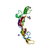 9dpmC  9dpnC  9dpoC  9dppC  9dpqC  9dprC  9dpsC  9dptC  9dpuC  9dpvC  9dpwC  9dpxC C: citing same article ( |
|---|---|
| Similar structure data |
-Links
 PDBj
PDBj


- Assembly
| Deposited unit | 
| ||||||||||||
|---|---|---|---|---|---|---|---|---|---|---|---|---|---|
| 1 |
| ||||||||||||
| Unit cell |
| ||||||||||||
| Components on special symmetry positions |
|
-Components
| #1: Protein | Mass: 12163.132 Da / Num. of mol.: 2 / Mutation: N357F, Y358F, A374T, Y409L, F411Y Source method: isolated from a genetically manipulated source Source: (gene. exp.) Homo sapiens (human) / Gene: BMP10 / Plasmid: pcdna3.1 / Cell line (production host): expi293 (Invitrogen) / Organ (production host): kidney / Production host: Homo sapiens (human) / Tissue (production host): embryonic / References: UniProt: O95393#2: Chemical | ChemComp-PO4 /   Mass: 94.971 Da / Num. of mol.: 13 / Source method: obtained synthetically / Formula: PO4 Mass: 94.971 Da / Num. of mol.: 13 / Source method: obtained synthetically / Formula: PO4#3: Chemical |   Mass: 22.990 Da / Num. of mol.: 2 / Source method: obtained synthetically / Formula: Na Mass: 22.990 Da / Num. of mol.: 2 / Source method: obtained synthetically / Formula: Na#4: Water | ChemComp-HOH / |  Mass: 18.015 Da / Num. of mol.: 144 / Source method: isolated from a natural source / Formula: H2O Mass: 18.015 Da / Num. of mol.: 144 / Source method: isolated from a natural source / Formula: H2OHas ligand of interest | N | Has protein modification | Y | |
|---|
-Experimental details
-Experiment
| Experiment | Method: X-RAY DIFFRACTION / Number of used crystals: 1 |
|---|
- Sample preparation
| Crystal | Density Matthews: 3.34 Å3/Da / Density % sol: 63.22 % |
|---|---|
| Crystal grow | Temperature: 293 K / Method: vapor diffusion, sitting drop / pH: 6.2 Details: 25% (v/v) 1,2-propanediol, 100 mM sodium phosphate dibasic/potassium phosphate monobasic pH 6.2, 10% (v/v) glycerol |
-Data collection
| Diffraction | Mean temperature: 100 K / Serial crystal experiment: N |
|---|---|
| Diffraction source | Source: SYNCHROTRON / Site: APS / Beamline: 24-ID-E / Wavelength: 0.97918 Å |
| Detector | Type: ADSC QUANTUM 315 / Detector: CCD / Date: Nov 28, 2022 |
| Radiation | Protocol: SINGLE WAVELENGTH / Monochromatic (M) / Laue (L): M / Scattering type: x-ray |
| Radiation wavelength | Wavelength: 0.97918 Å / Relative weight: 1 |
| Reflection | Resolution: 1.77→61.52 Å / Num. obs: 31025 / % possible obs: 98.8 % / Redundancy: 8.9 % / Biso Wilson estimate: 35.95 Å2 / CC1/2: 0.998 / Rmerge(I) obs: 0.078 / Rpim(I) all: 0.04 / Rrim(I) all: 0.088 / Χ2: 1.05 / Net I/σ(I): 12.2 |
| Reflection shell | Resolution: 1.77→1.87 Å / Redundancy: 8.9 % / Rmerge(I) obs: 2.426 / Mean I/σ(I) obs: 1 / Num. unique obs: 4391 / CC1/2: 0.531 / Rpim(I) all: 1.237 / Rrim(I) all: 2.727 / Χ2: 0.84 / % possible all: 97.2 |
- Processing
| Software |
| |||||||||||||||||||||||||||||||||||||||||||||||||||||||||||||||||||||||||||||||||||||||||||||||||||||||||
|---|---|---|---|---|---|---|---|---|---|---|---|---|---|---|---|---|---|---|---|---|---|---|---|---|---|---|---|---|---|---|---|---|---|---|---|---|---|---|---|---|---|---|---|---|---|---|---|---|---|---|---|---|---|---|---|---|---|---|---|---|---|---|---|---|---|---|---|---|---|---|---|---|---|---|---|---|---|---|---|---|---|---|---|---|---|---|---|---|---|---|---|---|---|---|---|---|---|---|---|---|---|---|---|---|---|---|
| Refinement | Method to determine structure: MOLECULAR REPLACEMENT / Resolution: 1.77→61.52 Å / SU ML: 0.2709 / Cross valid method: FREE R-VALUE / σ(F): 1.36 / Phase error: 37.6611 Stereochemistry target values: GeoStd + Monomer Library + CDL v1.2
| |||||||||||||||||||||||||||||||||||||||||||||||||||||||||||||||||||||||||||||||||||||||||||||||||||||||||
| Solvent computation | Shrinkage radii: 0.9 Å / VDW probe radii: 1.1 Å / Solvent model: FLAT BULK SOLVENT MODEL | |||||||||||||||||||||||||||||||||||||||||||||||||||||||||||||||||||||||||||||||||||||||||||||||||||||||||
| Displacement parameters | Biso mean: 60.29 Å2 | |||||||||||||||||||||||||||||||||||||||||||||||||||||||||||||||||||||||||||||||||||||||||||||||||||||||||
| Refinement step | Cycle: LAST / Resolution: 1.77→61.52 Å
| |||||||||||||||||||||||||||||||||||||||||||||||||||||||||||||||||||||||||||||||||||||||||||||||||||||||||
| Refine LS restraints |
| |||||||||||||||||||||||||||||||||||||||||||||||||||||||||||||||||||||||||||||||||||||||||||||||||||||||||
| LS refinement shell |
|
