 Movie
Movie Controller


+ Open data

- Basic information
| Entry | Database: PDB / ID: 9dnt | |||||||||
|---|---|---|---|---|---|---|---|---|---|---|
| Title | Cryo-EM structure of Tom1 (S. cerevisiae) | |||||||||
 Components Components | E3 ubiquitin-protein ligase TOM1 | |||||||||
 Keywords Keywords | TRANSFERASE / ubiquitin / ubiquitylation complex | |||||||||
| Function / homology |  Function and homology information Function and homology informationendonucleolytic cleavage in 5'-ETS of tricistronic rRNA transcript (SSU-rRNA, 5.8S rRNA, LSU-rRNA) / endonucleolytic cleavage to generate mature 5'-end of SSU-rRNA from (SSU-rRNA, 5.8S rRNA, LSU-rRNA) / HECT-type E3 ubiquitin transferase / nucleocytoplasmic transport / regulation of cell size / cleavage in ITS2 between 5.8S rRNA and LSU-rRNA of tricistronic rRNA transcript (SSU-rRNA, 5.8S rRNA, LSU-rRNA) / nucleus organization / Antigen processing: Ubiquitination & Proteasome degradation / mRNA transport / endonucleolytic cleavage in ITS1 to separate SSU-rRNA from 5.8S rRNA and LSU-rRNA from tricistronic rRNA transcript (SSU-rRNA, 5.8S rRNA, LSU-rRNA) ...endonucleolytic cleavage in 5'-ETS of tricistronic rRNA transcript (SSU-rRNA, 5.8S rRNA, LSU-rRNA) / endonucleolytic cleavage to generate mature 5'-end of SSU-rRNA from (SSU-rRNA, 5.8S rRNA, LSU-rRNA) / HECT-type E3 ubiquitin transferase / nucleocytoplasmic transport / regulation of cell size / cleavage in ITS2 between 5.8S rRNA and LSU-rRNA of tricistronic rRNA transcript (SSU-rRNA, 5.8S rRNA, LSU-rRNA) / nucleus organization / Antigen processing: Ubiquitination & Proteasome degradation / mRNA transport / endonucleolytic cleavage in ITS1 to separate SSU-rRNA from 5.8S rRNA and LSU-rRNA from tricistronic rRNA transcript (SSU-rRNA, 5.8S rRNA, LSU-rRNA) / Neutrophil degranulation / ubiquitin-protein transferase activity / ubiquitin protein ligase activity / mitotic cell cycle / ubiquitin-dependent protein catabolic process / protein ubiquitination / nucleolus / nucleus / cytoplasm Similarity search - Function | |||||||||
| Biological species |  | |||||||||
| Method | ELECTRON MICROSCOPY / single particle reconstruction / cryo EM / Resolution: 3 Å | |||||||||
 Authors Authors | Warner, K.M. / Hunkeler, M. / Baek, K. / Roy Burman, S.S. / Fischer, E.S. | |||||||||
| Funding support |  United States, 2items United States, 2items
| |||||||||
 Citation Citation | Journal: Cell Rep / Year: 2025 Title: Structural ubiquitin contributes to K48 linkage specificity of the HECT ligase Tom1. Authors: Katrina Warner / Moritz Hunkeler / Kheewoong Baek / Anna Schmoker / Shourya S Roy Burman / Daan Overwijn / Cyrus Jin / Katherine A Donovan / Eric S Fischer / Abstract: Homologous to E6AP C terminus (HECT) ubiquitin ligases play key roles in essential pathways such as DNA repair, cell cycle control, or protein quality control. Tom1 is one of five HECT ubiquitin E3 ...Homologous to E6AP C terminus (HECT) ubiquitin ligases play key roles in essential pathways such as DNA repair, cell cycle control, or protein quality control. Tom1 is one of five HECT ubiquitin E3 ligases in budding yeast S. cerevisiae and is prototypical for a ligase with pleiotropic functions such as ubiquitin chain amplification, orphan quality control, and DNA damage response. Structures of full-length HECT ligases, including the Tom1 ortholog HUWE1, have been reported, but how domains beyond the conserved catalytic module contribute to catalysis remains largely elusive. Here, through cryoelectron microscopy (cryo-EM) snapshots of Tom1 during an active ubiquitination cycle, we demonstrate that the extended domain architecture directly contributes to activity. We identify a Tom1-ubiquitin architecture during ubiquitination involving a non-canonical ubiquitin-binding site in the solenoid shape of Tom1. We demonstrate that this ubiquitin-binding site coordinates a structural ubiquitin contributing to the fidelity of K48 poly-ubiquitin chain assembly. | |||||||||
| History |
|
- Structure visualization
| Structure viewer | Molecule: MolmilJmol/JSmol |
|---|
- Downloads & links
-Download
| PDBx/mmCIF format | 9dnt.cif.gz | 965.9 KB | Display | PDBx/mmCIF format |
|---|---|---|---|---|
| PDB format | pdb9dnt.ent.gz | 796.6 KB | Display | PDB format |
| PDBx/mmJSON format | 9dnt.json.gz | Tree view | PDBx/mmJSON format | |
| Others |  Other downloads Other downloads |
-Validation report
| Arichive directory | https://data.pdbj.org/pub/pdb/validation_reports/dn/9dntftp://data.pdbj.org/pub/pdb/validation_reports/dn/9dnt | HTTPS FTP |
|---|
-Related structure data
| Related structure data |  47058MC  9dnsC M: map data used to model this data C: citing same article ( |
|---|---|
| Similar structure data |


-Links
 PDBj
PDBj


- Assembly
| Deposited unit | 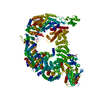
|
|---|---|
| 1 |
|
-Components
| #1: Protein | Mass: 377412.500 Da / Num. of mol.: 1 Source method: isolated from a genetically manipulated source Source: (gene. exp.) Gene: TOM1, SSR2, YDR457W, D8035.1 / Production host:  Trichoplusia ni (cabbage looper) Trichoplusia ni (cabbage looper)References: UniProt: Q03280, HECT-type E3 ubiquitin transferase |
|---|---|
| Has protein modification | N |
-Experimental details
-Experiment
| Experiment | Method: ELECTRON MICROSCOPY |
|---|---|
| EM experiment | Aggregation state: PARTICLE / 3D reconstruction method: single particle reconstruction |
- Sample preparation
| Component | Name: Full length structure, S. cerevisiae Tom1 / Type: COMPLEX / Details: Tom1 closed-ring conformation, stabilized / Entity ID: all / Source: RECOMBINANT | |||||||||||||||||||||||||
|---|---|---|---|---|---|---|---|---|---|---|---|---|---|---|---|---|---|---|---|---|---|---|---|---|---|---|
| Molecular weight | Value: 0.376 MDa / Experimental value: NO | |||||||||||||||||||||||||
| Source (natural) | Organism: | |||||||||||||||||||||||||
| Source (recombinant) | Organism: Trichoplusia ni (cabbage looper) | |||||||||||||||||||||||||
| Buffer solution | pH: 7.2 Details: 30 mM HEPES pH 7.2, 200 mM NaCl, 2 mM TCEP, 0.3 mM CHAPSO | |||||||||||||||||||||||||
| Buffer component |
| |||||||||||||||||||||||||
| Specimen | Conc.: 4.8 mg/ml / Embedding applied: NO / Shadowing applied: NO / Staining applied: NO / Vitrification applied: YES | |||||||||||||||||||||||||
| Specimen support | Details: 60 seconds / hold time: 10 seconds / Current: 15 mA Grid material: COPPER / Grid mesh size: 300 divisions/in. / Grid type: Quantifoil R1.2/1.3 | |||||||||||||||||||||||||
| Vitrification | Instrument: LEICA EM GP / Cryogen name: ETHANE / Humidity: 90 % / Chamber temperature: 283.15 K Details: CHAPSO detergent added to final conc. of 0.3 mM. Sample applied twice. |
- Electron microscopy imaging
| Experimental equipment |  Model: Titan Krios / Image courtesy: FEI Company |
|---|---|
| Microscopy | Model: TFS KRIOS |
| Electron gun | Electron source:  FIELD EMISSION GUN / Accelerating voltage: 300 kV / Illumination mode: FLOOD BEAM FIELD EMISSION GUN / Accelerating voltage: 300 kV / Illumination mode: FLOOD BEAM |
| Electron lens | Mode: BRIGHT FIELD / Nominal magnification: 105000 X / Nominal defocus max: 2200 nm / Nominal defocus min: 1100 nm / Cs: 2.7 mm / C2 aperture diameter: 50 µm / Alignment procedure: COMA FREE |
| Specimen holder | Cryogen: NITROGEN / Specimen holder model: FEI TITAN KRIOS AUTOGRID HOLDER |
| Image recording | Average exposure time: 2.8 sec. / Electron dose: 53.687 e/Å2 / Film or detector model: GATAN K3 BIOQUANTUM (6k x 4k) / Num. of grids imaged: 1 / Num. of real images: 11583 |
| EM imaging optics | Energyfilter name: GIF Bioquantum / Energyfilter slit width: 20 eV |
- Processing
| EM software |
| ||||||||||||||||||||||||||||||||||||||||||||
|---|---|---|---|---|---|---|---|---|---|---|---|---|---|---|---|---|---|---|---|---|---|---|---|---|---|---|---|---|---|---|---|---|---|---|---|---|---|---|---|---|---|---|---|---|---|
| CTF correction | Type: PHASE FLIPPING AND AMPLITUDE CORRECTION | ||||||||||||||||||||||||||||||||||||||||||||
| Particle selection | Num. of particles selected: 4216890 | ||||||||||||||||||||||||||||||||||||||||||||
| 3D reconstruction | Resolution: 3 Å / Resolution method: FSC 0.143 CUT-OFF / Num. of particles: 451216 / Symmetry type: POINT | ||||||||||||||||||||||||||||||||||||||||||||
| Atomic model building | B value: 121 | ||||||||||||||||||||||||||||||||||||||||||||
| Atomic model building | Source name: AlphaFold / Type: in silico model | ||||||||||||||||||||||||||||||||||||||||||||
| Refine LS restraints |
|
