 Movie
Movie Controller

[English] 日本語
 Yorodumi
- PDB-9dks: Crystal structure of CcbD C19S mutant complexed with single PCP d... -
+ Open data

- Basic information
| Entry | Database: PDB / ID: 9dks | |||||||||
|---|---|---|---|---|---|---|---|---|---|---|
| Title | Crystal structure of CcbD C19S mutant complexed with single PCP domain of CcbZ | |||||||||
 Components Components |
| |||||||||
 Keywords Keywords | BIOSYNTHETIC PROTEIN / complex / sugar / carrier protein | |||||||||
| Function / homology |  Function and homology information Function and homology information | |||||||||
| Biological species |  Streptomyces caelestis (bacteria) Streptomyces caelestis (bacteria) | |||||||||
| Method |  X-RAY DIFFRACTION / SYNCHROTRON / MOLECULAR REPLACEMENT / Resolution: 1.55 Å X-RAY DIFFRACTION / SYNCHROTRON / MOLECULAR REPLACEMENT / Resolution: 1.55 Å | |||||||||
 Authors Authors | Heberlig, G.W. / Podust, L.M. / Burkart, M.D. | |||||||||
| Funding support |  United States, 2items United States, 2items
| |||||||||
 Citation Citation | Journal: To Be Published Title: Crystal structure of CcbD complexed with PCP domain of CcbZ Authors: Heberlig, G.W. / Podust, L.M. / Burkart, M.D. | |||||||||
| History |
|
- Structure visualization
| Structure viewer | Molecule: MolmilJmol/JSmol |
|---|
- Downloads & links
-Download
| PDBx/mmCIF format | 9dks.cif.gz | 314.6 KB | Display | PDBx/mmCIF format |
|---|---|---|---|---|
| PDB format | pdb9dks.ent.gz | 246.5 KB | Display | PDB format |
| PDBx/mmJSON format | 9dks.json.gz | Tree view | PDBx/mmJSON format | |
| Others |  Other downloads Other downloads |
-Validation report
| Arichive directory | https://data.pdbj.org/pub/pdb/validation_reports/dk/9dksftp://data.pdbj.org/pub/pdb/validation_reports/dk/9dks | HTTPS FTP |
|---|
-Related structure data

-Links
 PDBj
PDBj
- Assembly
| Deposited unit | 
| ||||||||
|---|---|---|---|---|---|---|---|---|---|
| 1 |
| ||||||||
| Unit cell |
|
-Components
| #1: Protein | Mass: 39182.414 Da / Num. of mol.: 2 / Mutation: C19S Source method: isolated from a genetically manipulated source Source: (gene. exp.) Streptomyces caelestis (bacteria) / Gene: HDA41_002772 / Production host: #2: Protein | | Mass: 11201.400 Da / Num. of mol.: 1 Source method: isolated from a genetically manipulated source Source: (gene. exp.) Streptomyces caelestis (bacteria) / Gene: HDA41_002769 / Production host: #3: Chemical |   Mass: 102.046 Da / Num. of mol.: 2 / Source method: obtained synthetically / Formula: C3H2O4 Mass: 102.046 Da / Num. of mol.: 2 / Source method: obtained synthetically / Formula: C3H2O4#4: Chemical | ChemComp-PNS / | 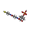  Mass: 358.348 Da / Num. of mol.: 1 / Source method: obtained synthetically / Formula: C11H23N2O7PS / Feature type: SUBJECT OF INVESTIGATION Mass: 358.348 Da / Num. of mol.: 1 / Source method: obtained synthetically / Formula: C11H23N2O7PS / Feature type: SUBJECT OF INVESTIGATION#5: Water | ChemComp-HOH / |  Mass: 18.015 Da / Num. of mol.: 660 / Source method: isolated from a natural source / Formula: H2O Mass: 18.015 Da / Num. of mol.: 660 / Source method: isolated from a natural source / Formula: H2OHas ligand of interest | Y | Has protein modification | N | |
|---|
-Experimental details
-Experiment
| Experiment | Method: X-RAY DIFFRACTION / Number of used crystals: 1 |
|---|
- Sample preparation
| Crystal | Density Matthews: 2.55 Å3/Da / Density % sol: 51.69 % |
|---|---|
| Crystal grow | Temperature: 295 K / Method: vapor diffusion, hanging drop / pH: 7 / Details: 0.2 M sodium malonate, 20% w/v PEG3350 |
-Data collection
| Diffraction | Mean temperature: 100 K / Serial crystal experiment: N |
|---|---|
| Diffraction source | Source: SYNCHROTRON / Site: ALS / Beamline: 8.3.1 / Wavelength: 1.11588 Å |
| Detector | Type: DECTRIS PILATUS3 S 6M / Detector: PIXEL / Date: Apr 20, 2022 |
| Radiation | Protocol: SINGLE WAVELENGTH / Monochromatic (M) / Laue (L): M / Scattering type: x-ray |
| Radiation wavelength | Wavelength: 1.11588 Å / Relative weight: 1 |
| Reflection | Resolution: 1.55→135.03 Å / Num. obs: 132094 / % possible obs: 100 % / Redundancy: 13 % / CC1/2: 1 / Rmerge(I) obs: 0.076 / Rpim(I) all: 0.032 / Rrim(I) all: 0.083 / Net I/σ(I): 18.7 |
| Reflection shell | Resolution: 1.55→1.58 Å / Redundancy: 12.9 % / Rmerge(I) obs: 1.973 / Mean I/σ(I) obs: 1.5 / Num. unique obs: 6507 / CC1/2: 0.649 / Rpim(I) all: 0.826 / Rrim(I) all: 2.141 / % possible all: 100 |
- Processing
| Software |
| |||||||||||||||||||||||||||||||||||||||||||||||||||||||||||||||||||||||||||||||||||||||||||||||||||||||||||||||||||||||||||||||||||||||||||||||||||||||||||
|---|---|---|---|---|---|---|---|---|---|---|---|---|---|---|---|---|---|---|---|---|---|---|---|---|---|---|---|---|---|---|---|---|---|---|---|---|---|---|---|---|---|---|---|---|---|---|---|---|---|---|---|---|---|---|---|---|---|---|---|---|---|---|---|---|---|---|---|---|---|---|---|---|---|---|---|---|---|---|---|---|---|---|---|---|---|---|---|---|---|---|---|---|---|---|---|---|---|---|---|---|---|---|---|---|---|---|---|---|---|---|---|---|---|---|---|---|---|---|---|---|---|---|---|---|---|---|---|---|---|---|---|---|---|---|---|---|---|---|---|---|---|---|---|---|---|---|---|---|---|---|---|---|---|---|---|---|
| Refinement | Method to determine structure: MOLECULAR REPLACEMENT / Resolution: 1.55→74.467 Å / Cor.coef. Fo:Fc: 0.971 / Cor.coef. Fo:Fc free: 0.961 / SU B: 1.863 / SU ML: 0.063 / Cross valid method: FREE R-VALUE / ESU R: 0.073 / ESU R Free: 0.075 Details: Hydrogens have been added in their riding positions
| |||||||||||||||||||||||||||||||||||||||||||||||||||||||||||||||||||||||||||||||||||||||||||||||||||||||||||||||||||||||||||||||||||||||||||||||||||||||||||
| Solvent computation | Ion probe radii: 0.8 Å / Shrinkage radii: 0.8 Å / VDW probe radii: 1.2 Å / Solvent model: MASK BULK SOLVENT | |||||||||||||||||||||||||||||||||||||||||||||||||||||||||||||||||||||||||||||||||||||||||||||||||||||||||||||||||||||||||||||||||||||||||||||||||||||||||||
| Displacement parameters | Biso mean: 25.25 Å2
| |||||||||||||||||||||||||||||||||||||||||||||||||||||||||||||||||||||||||||||||||||||||||||||||||||||||||||||||||||||||||||||||||||||||||||||||||||||||||||
| Refinement step | Cycle: LAST / Resolution: 1.55→74.467 Å
| |||||||||||||||||||||||||||||||||||||||||||||||||||||||||||||||||||||||||||||||||||||||||||||||||||||||||||||||||||||||||||||||||||||||||||||||||||||||||||
| Refine LS restraints |
| |||||||||||||||||||||||||||||||||||||||||||||||||||||||||||||||||||||||||||||||||||||||||||||||||||||||||||||||||||||||||||||||||||||||||||||||||||||||||||
| LS refinement shell |
|
