 Movie
Movie Controller


+ Open data

- Basic information
| Entry | Database: PDB / ID: 9djn | |||||||||
|---|---|---|---|---|---|---|---|---|---|---|
| Title | T4 Lysozyme K147H/T151H co-crystallized with Cu(II)-NTA | |||||||||
 Components Components | Endolysin | |||||||||
 Keywords Keywords | HYDROLASE / Hydrolase (O-Glycosyl) / double histidine mutation / dHis-Cu(II)-NTA motif / lysozyme | |||||||||
| Function / homology |  Function and homology information Function and homology informationviral release from host cell by cytolysis / peptidoglycan catabolic process / cell wall macromolecule catabolic process / lysozyme / lysozyme activity / host cell cytoplasm / defense response to bacterium Similarity search - Function | |||||||||
| Biological species |  Tequatrovirus T4 Tequatrovirus T4 | |||||||||
| Method |  X-RAY DIFFRACTION / SYNCHROTRON / MOLECULAR REPLACEMENT / Resolution: 1.26 Å X-RAY DIFFRACTION / SYNCHROTRON / MOLECULAR REPLACEMENT / Resolution: 1.26 Å | |||||||||
 Authors Authors | Besaw, J.E. / Ernst, O.P. | |||||||||
| Funding support |  Canada, 2items Canada, 2items
| |||||||||
 Citation Citation | Journal: Structure / Year: 2026 Title: Structural characterization of the Cu(II)-NTA spin label on alpha-helices by X-ray crystallography and electron paramagnetic resonance. Authors: Besaw, J.E. / Reichenwallner, J. / Chen, E.Y. / Hermet-Teesalu, P. / Tregubenko, A. / Kim, K. / Morizumi, T. / Ustav Jr., M. / Ernst, O.P. | |||||||||
| History |
|
- Structure visualization
| Structure viewer | Molecule: MolmilJmol/JSmol |
|---|
- Downloads & links
-Download
| PDBx/mmCIF format | 9djn.cif.gz | 91.4 KB | Display | PDBx/mmCIF format |
|---|---|---|---|---|
| PDB format | pdb9djn.ent.gz | 63.6 KB | Display | PDB format |
| PDBx/mmJSON format | 9djn.json.gz | Tree view | PDBx/mmJSON format | |
| Others |  Other downloads Other downloads |
-Validation report
| Arichive directory | https://data.pdbj.org/pub/pdb/validation_reports/dj/9djnftp://data.pdbj.org/pub/pdb/validation_reports/dj/9djn | HTTPS FTP |
|---|
-Related structure data
| Related structure data |  9d9jC  9djaC  9djbC  9djcC  9djdC  9djeC  9djfC  9djgC  9djhC  9djiC  9djjC  9djkC  9djlC  9djmC  9djoC  9djpC  9djqC  9djrC 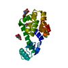 9djsC C: citing same article ( |
|---|---|
| Similar structure data |
-Links
 PDBj
PDBj


- Assembly
| Deposited unit | 
| ||||||||||||
|---|---|---|---|---|---|---|---|---|---|---|---|---|---|
| 1 |
| ||||||||||||
| Unit cell |
|
-Components
-Protein , 1 types, 1 molecules A
| #1: Protein | Mass: 18674.373 Da / Num. of mol.: 1 / Mutation: C54T, C97A, K147H, T151H Source method: isolated from a genetically manipulated source Source: (gene. exp.) Tequatrovirus T4 / Gene: e, T4Tp126 / Production host:  |
|---|
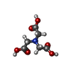
-Non-polymers , 8 types, 283 molecules 


| #2: Chemical | ChemComp-K /  Mass: 39.098 Da / Num. of mol.: 1 / Source method: obtained synthetically / Formula: K Mass: 39.098 Da / Num. of mol.: 1 / Source method: obtained synthetically / Formula: K | ||||||||||
|---|---|---|---|---|---|---|---|---|---|---|---|
| #3: Chemical | ChemComp-PO4 /  Mass: 94.971 Da / Num. of mol.: 1 / Source method: obtained synthetically / Formula: PO4 Mass: 94.971 Da / Num. of mol.: 1 / Source method: obtained synthetically / Formula: PO4 | ||||||||||
| #4: Chemical |  Mass: 35.453 Da / Num. of mol.: 3 / Source method: obtained synthetically / Formula: Cl Mass: 35.453 Da / Num. of mol.: 3 / Source method: obtained synthetically / Formula: Cl#5: Chemical | ChemComp-CU / |  Mass: 63.546 Da / Num. of mol.: 1 / Source method: obtained synthetically / Formula: Cu / Feature type: SUBJECT OF INVESTIGATION Mass: 63.546 Da / Num. of mol.: 1 / Source method: obtained synthetically / Formula: Cu / Feature type: SUBJECT OF INVESTIGATION#6: Chemical | ChemComp-NTA / |  Mass: 191.139 Da / Num. of mol.: 1 / Source method: obtained synthetically / Formula: C6H9NO6 / Feature type: SUBJECT OF INVESTIGATION Mass: 191.139 Da / Num. of mol.: 1 / Source method: obtained synthetically / Formula: C6H9NO6 / Feature type: SUBJECT OF INVESTIGATION#7: Chemical | ChemComp-NA / |  Mass: 22.990 Da / Num. of mol.: 1 / Source method: obtained synthetically / Formula: Na Mass: 22.990 Da / Num. of mol.: 1 / Source method: obtained synthetically / Formula: Na#8: Chemical | ChemComp-HEZ / |  Mass: 118.174 Da / Num. of mol.: 1 / Source method: obtained synthetically / Formula: C6H14O2 Mass: 118.174 Da / Num. of mol.: 1 / Source method: obtained synthetically / Formula: C6H14O2#9: Water | ChemComp-HOH / | Mass: 18.015 Da / Num. of mol.: 274 / Source method: isolated from a natural source / Formula: H2O |
-Details
| Has ligand of interest | Y |
|---|---|
| Has protein modification | N |
-Experimental details
-Experiment
| Experiment | Method: X-RAY DIFFRACTION / Number of used crystals: 1 |
|---|
- Sample preparation
| Crystal | Density Matthews: 2.59 Å3/Da / Density % sol: 52.54 % |
|---|---|
| Crystal grow | Temperature: 293 K / Method: vapor diffusion, hanging drop / pH: 6.6 Details: Protein: 0.33 mM T4 lysozyme mutant, 3.3 mM Cu(II)-NTA. Precipitant: 2.2 M NaH2PO4/K2HPO4, pH 6.6, 150 mM NaCl, 100 mM 1,6-hexanediol, 3% 2-propanol |
-Data collection
| Diffraction | Mean temperature: 100 K / Serial crystal experiment: N |
|---|---|
| Diffraction source | Source: SYNCHROTRON / Site: NSLS-II  / Beamline: 17-ID-2 / Wavelength: 0.9794 Å / Beamline: 17-ID-2 / Wavelength: 0.9794 Å |
| Detector | Type: DECTRIS EIGER X 16M / Detector: PIXEL / Date: Nov 21, 2022 |
| Radiation | Protocol: SINGLE WAVELENGTH / Monochromatic (M) / Laue (L): M / Scattering type: x-ray |
| Radiation wavelength | Wavelength: 0.9794 Å / Relative weight: 1 |
| Reflection | Resolution: 1.26→28.33 Å / Num. obs: 53137 / % possible obs: 99.8 % / Redundancy: 10.9 % / Biso Wilson estimate: 11.35 Å2 / CC1/2: 0.999 / Net I/σ(I): 15.9 |
| Reflection shell | Resolution: 1.26→1.29 Å / Redundancy: 9.9 % / Mean I/σ(I) obs: 2.8 / Num. unique obs: 3759 / CC1/2: 0.883 / % possible all: 97.2 |
- Processing
| Software |
| |||||||||||||||||||||||||||||||||||||||||||||||||||||||||||||||||||||||||||||||||||||||||||||||||||||||||
|---|---|---|---|---|---|---|---|---|---|---|---|---|---|---|---|---|---|---|---|---|---|---|---|---|---|---|---|---|---|---|---|---|---|---|---|---|---|---|---|---|---|---|---|---|---|---|---|---|---|---|---|---|---|---|---|---|---|---|---|---|---|---|---|---|---|---|---|---|---|---|---|---|---|---|---|---|---|---|---|---|---|---|---|---|---|---|---|---|---|---|---|---|---|---|---|---|---|---|---|---|---|---|---|---|---|---|
| Refinement | Method to determine structure: MOLECULAR REPLACEMENT / Resolution: 1.26→27 Å / SU ML: 0.0849 / Cross valid method: FREE R-VALUE / σ(F): 1.36 / Phase error: 14.9942 Stereochemistry target values: GeoStd + Monomer Library + CDL v1.2
| |||||||||||||||||||||||||||||||||||||||||||||||||||||||||||||||||||||||||||||||||||||||||||||||||||||||||
| Solvent computation | Shrinkage radii: 0.9 Å / VDW probe radii: 1.1 Å / Solvent model: FLAT BULK SOLVENT MODEL | |||||||||||||||||||||||||||||||||||||||||||||||||||||||||||||||||||||||||||||||||||||||||||||||||||||||||
| Displacement parameters | Biso mean: 15.47 Å2 | |||||||||||||||||||||||||||||||||||||||||||||||||||||||||||||||||||||||||||||||||||||||||||||||||||||||||
| Refinement step | Cycle: LAST / Resolution: 1.26→27 Å
| |||||||||||||||||||||||||||||||||||||||||||||||||||||||||||||||||||||||||||||||||||||||||||||||||||||||||
| Refine LS restraints |
| |||||||||||||||||||||||||||||||||||||||||||||||||||||||||||||||||||||||||||||||||||||||||||||||||||||||||
| LS refinement shell |
|
