[English] 日本語
Yorodumi
- PDB-9dib: Rous sarcoma virus frameshifting pseudoknot RNA -

+
Open data


ID or keywords:

Loading...

-
Basic information

Entry
Database: PDB / ID: 9dib
TitleRous sarcoma virus frameshifting pseudoknot RNA
Componentsframeshifting pseudoknot RNA
KeywordsRNA / pseudoknot / retroviral RNA / frameshifting / translation regulation
Function / homologyIRIDIUM HEXAMMINE ION / : / : / RNA / RNA (> 10) / RNA (> 100)
Function and homology information
Biological speciesRous sarcoma virus - Prague C
MethodX-RAY DIFFRACTION / SYNCHROTRON / MOLECULAR REPLACEMENT / Resolution: 2.68 Å
AuthorsJones, C.P. / Ferre-D'Amare, A.R.
Funding support United States, 1items
OrganizationGrant numberCountry
National Institutes of Health/National Heart, Lung, and Blood Institute (NIH/NHLBI) United States
CitationJournal: Proc Natl Acad Sci U S A / Year: 2025
Title: Structural switching dynamically controls the doubly pseudoknotted Rous sarcoma virus-programmed ribosomal frameshifting element.
Authors: Christopher P Jones / Adrian R Ferré-D'Amaré /
Abstract: A hallmark of retrovirus replication is the translation of two different polyproteins from one RNA through programmed -1 frameshifting. This is a mechanism in which the actively translating ribosome ...A hallmark of retrovirus replication is the translation of two different polyproteins from one RNA through programmed -1 frameshifting. This is a mechanism in which the actively translating ribosome is induced to slip in the 5' direction at a defined codon and then continues translating in the new reading frame. Programmed frameshifting controls the stoichiometry of viral proteins and is therefore under stringent evolutionary selection. Forty years ago, the first frameshifting stimulatory element was discovered in the Rous sarcoma virus. The ~120 nt RNA segment was predicted to contain a pseudoknot, but its 3D structure has remained elusive. Now, we have determined cryoEM and X-ray crystallographic structures of this classic retroviral element, finding that it adopts a butterfly-like double-pseudoknot fold. One "wing" contains a dynamic pyrimidine-rich helix, observed crystallographically in two conformations and in a third conformation via cryoEM. The other wing encompasses the predicted pseudoknot, which interacts with a second unexpected pseudoknot through a toggle residue, A2546. This key purine switches conformations between structural states and tunes the stability of interacting residues in the two wings. We find that its mutation can modulate frameshifting by as much as 50-fold, likely by altering the relative abundance of different structural states in the conformational ensemble of the RNA. Taken together, our structure-function analyses reveal how a dynamic double pseudoknot junction stimulates frameshifting by taking advantage of conformational heterogeneity, supporting a multistate model in which high Shannon entropy enhances frameshifting efficiency.
History
DepositionSep 5, 2024Deposition site: RCSB / Processing site: RCSB
Revision 1.0Apr 9, 2025Provider: repository / Type: Initial release
Revision 1.1Apr 16, 2025Group: Database references / Category: citation / citation_author
Item: _citation.journal_volume / _citation.page_first ..._citation.journal_volume / _citation.page_first / _citation.page_last / _citation.pdbx_database_id_DOI / _citation.pdbx_database_id_PubMed / _citation.title / _citation_author.identifier_ORCID

-
Structure visualization

Structure viewerMolecule:
MolmilJmol/JSmol

Downloads & links

-
Assembly

Deposited unit
A: frameshifting pseudoknot RNA
B: frameshifting pseudoknot RNA
hetero molecules


Theoretical massNumber of molelcules
Total (without water)76,95229
Polymers71,3602
Non-polymers5,59227
Water25214
1


  • Idetical with deposited unit
  • defined by author
  • Evidence: gel filtration, Dimerized in folded RNA solution in an ~1:5 ratio of dimer:monomer
TypeNameSymmetry operationNumber
identity operation1_555x,y,z1
Buried area8130 Å2
ΔGint-46 kcal/mol
Surface area33720 Å2
Unit cell
Length a, b, c (Å)43.763, 68.246, 209.563
Angle α, β, γ (deg.)90.00, 90.00, 90.00
Int Tables number19
Space group name H-MP212121

-
Components

#1: RNA chain frameshifting pseudoknot RNA


Mass: 35680.164 Da / Num. of mol.: 2
Source method: isolated from a genetically manipulated source
Details: Purified from in vitro transcription via gel electrophoresis and elution then refolded (see doi: 10.15252/embj.201489209 for general RNA prep details)
Source: (gene. exp.) Rous sarcoma virus - Prague C
Production host: in vitro transcription vector pT7-TP(deltai) (others)
References: GenBank: 210171
#2: Chemical
ChemComp-IRI / IRIDIUM HEXAMMINE ION


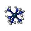
Mass: 294.400 Da / Num. of mol.: 18 / Source method: obtained synthetically / Formula: H18IrN6
#3: Chemical
ChemComp-MG / MAGNESIUM ION


Mass: 24.305 Da / Num. of mol.: 4 / Source method: obtained synthetically / Formula: Mg
#4: Chemical
ChemComp-K / POTASSIUM ION


Mass: 39.098 Da / Num. of mol.: 5 / Source method: obtained synthetically / Formula: K
#5: Water ChemComp-HOH / water


Mass: 18.015 Da / Num. of mol.: 14 / Source method: isolated from a natural source / Formula: H2O
Has ligand of interestN
Has protein modificationN

-
Experimental details

-
Experiment

ExperimentMethod: X-RAY DIFFRACTION / Number of used crystals: 1

-
Sample preparation

CrystalDensity Matthews: 2.19 Å3/Da / Density % sol: 43.91 %
Crystal growTemperature: 294 K / Method: vapor diffusion, hanging drop
Details: 20% w/v PEG3350, 0.25 M sodium chloride, 0.1 M Tris-HCl, pH 8.5

-
Data collection

DiffractionMean temperature: 100 K / Serial crystal experiment: N
Diffraction sourceSource: SYNCHROTRON / Site: ALS / Beamline: 5.0.1 / Wavelength: 0.977 Å
DetectorType: DECTRIS PILATUS 2M / Detector: PIXEL / Date: May 19, 2021
RadiationProtocol: SINGLE WAVELENGTH / Monochromatic (M) / Laue (L): M / Scattering type: x-ray
Radiation wavelengthWavelength: 0.977 Å / Relative weight: 1
ReflectionResolution: 2.68→48.82 Å / Num. obs: 18350 / % possible obs: 99.4 % / Redundancy: 12.9 % / CC1/2: 1 / Rmerge(I) obs: 0.0935 / Net I/σ(I): 22.5
Reflection shellResolution: 2.68→2.776 Å / Rmerge(I) obs: 1.47 / Mean I/σ(I) obs: 1.62 / Num. unique obs: 1815 / CC1/2: 0.776

-
Processing

Software
NameVersionClassification
PHENIX(1.19.2_4158: ???)refinement
Aimlessdata scaling
XDSdata reduction
AutoSolphasing
RefinementMethod to determine structure: MOLECULAR REPLACEMENT / Resolution: 2.68→48.82 Å / SU ML: 0.47 / Cross valid method: FREE R-VALUE / σ(F): 1.34 / Phase error: 33.05 / Stereochemistry target values: ML
RfactorNum. reflection% reflection
Rfree0.2679 3068 9.05 %
Rwork0.2057 --
obs0.2113 18350 99.48 %
Solvent computationShrinkage radii: 0.9 Å / VDW probe radii: 1.11 Å / Solvent model: FLAT BULK SOLVENT MODEL
Refinement stepCycle: LAST / Resolution: 2.68→48.82 Å
ProteinNucleic acidLigandSolventTotal
Num. atoms0 4190 135 14 4339
Refine LS restraints
Refine-IDTypeDev idealNumber
X-RAY DIFFRACTIONf_bond_d0.0064842
X-RAY DIFFRACTIONf_angle_d1.147638
X-RAY DIFFRACTIONf_dihedral_angle_d13.9332383
X-RAY DIFFRACTIONf_chiral_restr0.0521000
X-RAY DIFFRACTIONf_plane_restr0.008198
LS refinement shell
Resolution (Å)Rfactor RfreeNum. reflection RfreeRfactor RworkNum. reflection RworkRefine-ID% reflection obs (%)
2.68-2.720.37621400.37641450X-RAY DIFFRACTION100
2.72-2.770.39821410.36341377X-RAY DIFFRACTION100
2.77-2.810.4121420.35431423X-RAY DIFFRACTION100
2.81-2.870.40941380.33611392X-RAY DIFFRACTION100
2.87-2.920.43731410.331424X-RAY DIFFRACTION99
2.92-2.980.43171350.3261360X-RAY DIFFRACTION100
2.98-3.040.32061410.27631390X-RAY DIFFRACTION99
3.05-3.120.39581450.24711448X-RAY DIFFRACTION98
3.12-3.190.26071340.22951318X-RAY DIFFRACTION98
3.19-3.280.28271330.19471403X-RAY DIFFRACTION99
3.28-3.380.28761430.19691434X-RAY DIFFRACTION99
3.38-3.490.27631390.18431367X-RAY DIFFRACTION99
3.49-3.610.22371360.18421421X-RAY DIFFRACTION99
3.61-3.750.22341370.1721361X-RAY DIFFRACTION99
3.75-3.930.20571450.16021399X-RAY DIFFRACTION99
3.93-4.130.21721400.14971404X-RAY DIFFRACTION100
4.13-4.390.23421450.16551431X-RAY DIFFRACTION100
4.39-4.730.23731420.16361384X-RAY DIFFRACTION100
4.73-5.20.24971370.17971400X-RAY DIFFRACTION100
5.21-5.950.20351390.18931406X-RAY DIFFRACTION100
5.96-7.490.26341380.1981428X-RAY DIFFRACTION100
7.5-48.820.28821370.23491412X-RAY DIFFRACTION100
Refinement TLS params.

Method: refined / Refine-ID: X-RAY DIFFRACTION

IDL112)L122)L132)L222)L232)L332)S11 (Å °)S12 (Å °)S13 (Å °)S21 (Å °)S22 (Å °)S23 (Å °)S31 (Å °)S32 (Å °)S33 (Å °)T112)T122)T132)T222)T232)T332)Origin x (Å)Origin y (Å)Origin z (Å)
16.0737-3.2322.32344.7079-4.5416.1459-0.791-1.0634-0.12240.570.3237-0.16370.8441-1.17930.43171.2598-0.07910.14621.2061-0.18660.636673.191366.254284.2707
20.0647-0.1445-1.40922.00110.46388.6202-0.24040.2648-0.0247-0.39190.2662-0.01051.14-0.2744-0.01540.88360.0565-0.03860.8488-0.12290.632754.324163.4865108.4437
33.05641.9139-0.37214.8488-2.07470.93440.5123-0.1326-0.24590.998-0.19890.454-0.2509-0.001-0.17580.94210.0014-0.01540.8125-0.11150.572349.066959.9221132.9475
4-0.34130.0635-1.1752-0.28261.36227.6219-0.04750.0262-0.1677-0.05220.03490.00530.19740.5909-0.10641.00550.0788-0.00741.1414-0.1290.636763.729566.5456102.0617
56.35424.4861.93328.11627.80659.48220.4123-0.5798-0.21511.13970.5222-1.13610.05480.8009-0.77280.75170.1154-0.11891.0149-0.18680.670150.464652.948876.9191
64.29995.9643.14968.46852.54985.01010.00420.7265-0.74930.40070.5637-0.32310.0334-0.0773-0.42770.46680.04270.01890.4918-0.03340.444249.240352.653358.7152
73.65971.15731.17691.607-0.64980.99040.28810.6663-0.2616-0.35970.1113-0.26280.0566-0.0258-0.29070.46640.17030.07630.5905-0.02090.399270.180560.816638.6239
85.06386.72983.64049.02744.85226.0938-0.18391.2851-2.2726-0.67732.0766-3.79881.15170.8784-0.52461.2622-0.00520.00191.2599-0.2591.141869.12455.773726.0491
93.9049-0.2240.6253.261-1.99393.55570.5170.7403-0.1528-0.3687-0.19460.223-0.24360.0942-0.31090.38650.0575-0.01580.4502-0.04410.256950.759952.04443.5223
100.18340.981.18245.0722.65136.93990.373-0.92990.01121.59140.5517-0.65711.00970.7351-0.96740.8870.1979-0.10521.1761-0.10580.690350.598253.26777.1815
110.9731-1.3664-0.48191.48931.00471.54470.4234-1.26121.15260.73960.21140.0341-0.065-0.6904-0.40620.6439-0.05720.16171.0315-0.23320.882152.621374.984864.1877
123.73263.57514.78635.12123.29446.97620.28570.4369-0.31240.11110.08-0.4090.36130.2373-0.34190.30990.0510.00540.39490.13430.346977.248662.267956.1369
Refinement TLS group
IDRefine-IDRefine TLS-IDSelection details
1X-RAY DIFFRACTION1chain 'A' and (resid 1 through 10 )
2X-RAY DIFFRACTION2chain 'A' and (resid 11 through 34 )
3X-RAY DIFFRACTION3chain 'A' and (resid 35 through 55 )
4X-RAY DIFFRACTION4chain 'A' and (resid 56 through 111 )
5X-RAY DIFFRACTION5chain 'B' and (resid 1 through 10 )
6X-RAY DIFFRACTION6chain 'B' and (resid 11 through 20 )
7X-RAY DIFFRACTION7chain 'B' and (resid 21 through 40 )
8X-RAY DIFFRACTION8chain 'B' and (resid 41 through 52 )
9X-RAY DIFFRACTION9chain 'B' and (resid 53 through 72 )
10X-RAY DIFFRACTION10chain 'B' and (resid 73 through 87 )
11X-RAY DIFFRACTION11chain 'B' and (resid 88 through 102 )
12X-RAY DIFFRACTION12(chain 'I' and resid 18)

+
About Yorodumi

-
News

-
Feb 9, 2022. New format data for meta-information of EMDB entries

New format data for meta-information of EMDB entries

  • Version 3 of the EMDB header file is now the official format.
  • The previous official version 1.9 will be removed from the archive.

Related info.:EMDB header

External links:wwPDB to switch to version 3 of the EMDB data model

-
Aug 12, 2020. Covid-19 info

Covid-19 info

URL: https://pdbj.org/emnavi/covid19.php

New page: Covid-19 featured information page in EM Navigator.

Related info.:Covid-19 info / Mar 5, 2020. Novel coronavirus structure data

+
Mar 5, 2020. Novel coronavirus structure data

Novel coronavirus structure data

Related info.:Yorodumi Speices / Aug 12, 2020. Covid-19 info

External links:COVID-19 featured content - PDBj / Molecule of the Month (242):Coronavirus Proteases

+
Jan 31, 2019. EMDB accession codes are about to change! (news from PDBe EMDB page)

EMDB accession codes are about to change! (news from PDBe EMDB page)

  • The allocation of 4 digits for EMDB accession codes will soon come to an end. Whilst these codes will remain in use, new EMDB accession codes will include an additional digit and will expand incrementally as the available range of codes is exhausted. The current 4-digit format prefixed with “EMD-” (i.e. EMD-XXXX) will advance to a 5-digit format (i.e. EMD-XXXXX), and so on. It is currently estimated that the 4-digit codes will be depleted around Spring 2019, at which point the 5-digit format will come into force.
  • The EM Navigator/Yorodumi systems omit the EMD- prefix.

Related info.:Q: What is EMD? / ID/Accession-code notation in Yorodumi/EM Navigator

External links:EMDB Accession Codes are Changing Soon! / Contact to PDBj

+
Jul 12, 2017. Major update of PDB

Major update of PDB

  • wwPDB released updated PDB data conforming to the new PDBx/mmCIF dictionary.
  • This is a major update changing the version number from 4 to 5, and with Remediation, in which all the entries are updated.
  • In this update, many items about electron microscopy experimental information are reorganized (e.g. em_software).
  • Now, EM Navigator and Yorodumi are based on the updated data.

External links:wwPDB Remediation / Enriched Model Files Conforming to OneDep Data Standards Now Available in the PDB FTP Archive

-
Yorodumi

Thousand views of thousand structures

  • Yorodumi is a browser for structure data from EMDB, PDB, SASBDB, etc.
  • This page is also the successor to EM Navigator detail page, and also detail information page/front-end page for Omokage search.
  • The word "yorodu" (or yorozu) is an old Japanese word meaning "ten thousand". "mi" (miru) is to see.

Related info.:EMDB / PDB / SASBDB / Comparison of 3 databanks / Yorodumi Search / Aug 31, 2016. New EM Navigator & Yorodumi / Yorodumi Papers / Jmol/JSmol / Function and homology information / Changes in new EM Navigator and Yorodumi

Read more