 Movie
Movie Controller

[English] 日本語
 Yorodumi
- PDB-9dfu: X-ray crystal structure of the second Viperin-like enzyme from T.... -
+ Open data

- Basic information
| Entry | Database: PDB / ID: 9dfu | ||||||
|---|---|---|---|---|---|---|---|
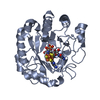
| Title | X-ray crystal structure of the second Viperin-like enzyme from T. virens variant F40H with bound CTP and SAM | ||||||
 Components Components | Radical SAM core domain-containing protein | ||||||
 Keywords Keywords | ANTIVIRAL PROTEIN / viperin-like enzyme / CTP / SAM / 4Fe-4S cluster / ddhCTP | ||||||
| Function / homology |  Function and homology information Function and homology informationcatalytic activity / 4 iron, 4 sulfur cluster binding / defense response to virus / nucleotide binding / mitochondrion / metal ion binding Similarity search - Function | ||||||
| Biological species |  Trichoderma virens (fungus) Trichoderma virens (fungus) | ||||||
| Method |  X-RAY DIFFRACTION / SYNCHROTRON / MOLECULAR REPLACEMENT / Resolution: 2.3 Å X-RAY DIFFRACTION / SYNCHROTRON / MOLECULAR REPLACEMENT / Resolution: 2.3 Å | ||||||
 Authors Authors | Lachowicz, J.C. / Bonanno, J.B. / Grove, T.L. | ||||||
| Funding support |  United States, 1items United States, 1items
| ||||||
 Citation Citation | Journal: Structure / Year: 2025 Title: Structural insights from active site variants and beta-8 loop interactions in viperin-like enzymes. Authors: Lachowicz, J.C. / Grudman, S. / Bonanno, J.B. / Fiser, A. / Grove, T.L. | ||||||
| History |
|
- Structure visualization
| Structure viewer | Molecule: MolmilJmol/JSmol |
|---|
- Downloads & links
-Download
| PDBx/mmCIF format | 9dfu.cif.gz | 78.5 KB | Display | PDBx/mmCIF format |
|---|---|---|---|---|
| PDB format | pdb9dfu.ent.gz | 54.8 KB | Display | PDB format |
| PDBx/mmJSON format | 9dfu.json.gz | Tree view | PDBx/mmJSON format | |
| Others |  Other downloads Other downloads |
-Validation report
| Summary document | 9dfu_validation.pdf.gz | 1.6 MB | Display | wwPDB validaton report |
|---|---|---|---|---|
| Full document | 9dfu_full_validation.pdf.gz | 1.6 MB | Display | |
| Data in XML | 9dfu_validation.xml.gz | 15.4 KB | Display | |
| Data in CIF | 9dfu_validation.cif.gz | 20 KB | Display | |
| Arichive directory | https://data.pdbj.org/pub/pdb/validation_reports/df/9dfuftp://data.pdbj.org/pub/pdb/validation_reports/df/9dfu | HTTPS FTP |
-Related structure data


-Links
 PDBj
PDBj

- Assembly
| Deposited unit | 
| ||||||||
|---|---|---|---|---|---|---|---|---|---|
| 1 |
| ||||||||
| Unit cell |
|
-Components
-Protein , 1 types, 1 molecules A
| #1: Protein | Mass: 37369.562 Da / Num. of mol.: 1 / Mutation: F40H Source method: isolated from a genetically manipulated source Source: (gene. exp.) Trichoderma virens (fungus) / Strain: Gv29-8 / Gene: TRIVIDRAFT_46896 / Production host:  |
|---|
-Non-polymers , 5 types, 95 molecules 


| #2: Chemical | ChemComp-CTP /  Mass: 483.156 Da / Num. of mol.: 1 / Source method: obtained synthetically / Formula: C9H16N3O14P3 / Feature type: SUBJECT OF INVESTIGATION Mass: 483.156 Da / Num. of mol.: 1 / Source method: obtained synthetically / Formula: C9H16N3O14P3 / Feature type: SUBJECT OF INVESTIGATION |
|---|---|
| #3: Chemical | ChemComp-SF4 /  Mass: 351.640 Da / Num. of mol.: 1 / Source method: isolated from a natural source / Formula: Fe4S4 / Feature type: SUBJECT OF INVESTIGATION Mass: 351.640 Da / Num. of mol.: 1 / Source method: isolated from a natural source / Formula: Fe4S4 / Feature type: SUBJECT OF INVESTIGATION |
| #4: Chemical | ChemComp-SAM /  Mass: 398.437 Da / Num. of mol.: 1 / Source method: obtained synthetically / Formula: C15H22N6O5S / Feature type: SUBJECT OF INVESTIGATION Mass: 398.437 Da / Num. of mol.: 1 / Source method: obtained synthetically / Formula: C15H22N6O5S / Feature type: SUBJECT OF INVESTIGATION |
| #5: Chemical | ChemComp-CL /  Mass: 35.453 Da / Num. of mol.: 1 / Source method: obtained synthetically / Formula: Cl Mass: 35.453 Da / Num. of mol.: 1 / Source method: obtained synthetically / Formula: Cl |
| #6: Water | ChemComp-HOH / Mass: 18.015 Da / Num. of mol.: 91 / Source method: isolated from a natural source / Formula: H2O |
-Details
| Has ligand of interest | Y |
|---|---|
| Has protein modification | N |
-Experimental details
-Experiment
| Experiment | Method: X-RAY DIFFRACTION / Number of used crystals: 1 |
|---|
- Sample preparation
| Crystal | Density Matthews: 2.12 Å3/Da / Density % sol: 42.1 % |
|---|---|
| Crystal grow | Temperature: 298 K / Method: vapor diffusion, sitting drop / pH: 7.5 / Details: 0.1 M HEPES (pH 7.5), 25% (w/v) PEG 3350 |
-Data collection
| Diffraction | Mean temperature: 100 K / Serial crystal experiment: N |
|---|---|
| Diffraction source | Source: SYNCHROTRON / Site: NSLS-II / Beamline: 17-ID-2 / Wavelength: 0.9201 Å |
| Detector | Type: DECTRIS EIGER X 16M / Detector: PIXEL / Date: Dec 13, 2023 |
| Radiation | Protocol: SINGLE WAVELENGTH / Monochromatic (M) / Laue (L): M / Scattering type: x-ray |
| Radiation wavelength | Wavelength: 0.9201 Å / Relative weight: 1 |
| Reflection | Resolution: 2.3→33.4 Å / Num. obs: 14839 / % possible obs: 99.7 % / Redundancy: 5.1 % / CC1/2: 0.979 / Rmerge(I) obs: 0.217 / Rpim(I) all: 0.154 / Rrim(I) all: 0.268 / Net I/σ(I): 5.3 |
| Reflection shell | Resolution: 2.3→2.38 Å / Rmerge(I) obs: 0.799 / Mean I/σ(I) obs: 2 / Num. unique obs: 1427 / CC1/2: 0.57 / Rpim(I) all: 0.553 / Rrim(I) all: 0.976 |
- Processing
| Software |
| ||||||||||||||||||||||||||||||||||||||||||
|---|---|---|---|---|---|---|---|---|---|---|---|---|---|---|---|---|---|---|---|---|---|---|---|---|---|---|---|---|---|---|---|---|---|---|---|---|---|---|---|---|---|---|---|
| Refinement | Method to determine structure: MOLECULAR REPLACEMENT / Resolution: 2.3→19.66 Å / SU ML: 0.3 / Cross valid method: FREE R-VALUE / σ(F): 1.34 / Phase error: 26.6 / Stereochemistry target values: ML
| ||||||||||||||||||||||||||||||||||||||||||
| Solvent computation | Shrinkage radii: 0.9 Å / VDW probe radii: 1.1 Å / Solvent model: FLAT BULK SOLVENT MODEL | ||||||||||||||||||||||||||||||||||||||||||
| Refinement step | Cycle: LAST / Resolution: 2.3→19.66 Å
| ||||||||||||||||||||||||||||||||||||||||||
| Refine LS restraints |
| ||||||||||||||||||||||||||||||||||||||||||
| LS refinement shell |
|
