 Movie
Movie Controller


+ Open data

- Basic information
| Entry | Database: PDB / ID: 9db6 | ||||||
|---|---|---|---|---|---|---|---|
| Title | Sialidase682 co-crystallized with inhibitor DANA | ||||||
 Components Components | BNR/Asp-box repeat protein | ||||||
 Keywords Keywords | HYDROLASE / Sialidase / sialic acid inhibitor complex | ||||||
| Function / homology | 2-DEOXY-2,3-DEHYDRO-N-ACETYL-NEURAMINIC ACID / :  Function and homology information Function and homology information | ||||||
| Biological species |  Bacteroides finegoldii DSM 17565 (bacteria) Bacteroides finegoldii DSM 17565 (bacteria) | ||||||
| Method |  X-RAY DIFFRACTION / SYNCHROTRON / MOLECULAR REPLACEMENT / Resolution: 2.1 Å X-RAY DIFFRACTION / SYNCHROTRON / MOLECULAR REPLACEMENT / Resolution: 2.1 Å | ||||||
 Authors Authors | Young, M.A. / Rees, S.D. / Chang, G. | ||||||
| Funding support | 1items
| ||||||
 Citation Citation | Journal: To Be Published Title: Sialidase682 co-crystallized with inhibitor DANA Authors: Young, M.A. / Rees, S.D. / Chang, G. | ||||||
| History |
|
- Structure visualization
| Structure viewer | Molecule: MolmilJmol/JSmol |
|---|
- Downloads & links
-Download
| PDBx/mmCIF format | 9db6.cif.gz | 915.3 KB | Display | PDBx/mmCIF format |
|---|---|---|---|---|
| PDB format | pdb9db6.ent.gz | 650 KB | Display | PDB format |
| PDBx/mmJSON format | 9db6.json.gz | Tree view | PDBx/mmJSON format | |
| Others |  Other downloads Other downloads |
-Validation report
| Arichive directory | https://data.pdbj.org/pub/pdb/validation_reports/db/9db6ftp://data.pdbj.org/pub/pdb/validation_reports/db/9db6 | HTTPS FTP |
|---|
-Related structure data
| Similar structure data |
|---|
-Links
 PDBj
PDBj- Assembly
| Deposited unit | 
| ||||||||||||
|---|---|---|---|---|---|---|---|---|---|---|---|---|---|
| 1 |
| ||||||||||||
| Unit cell |
|
-Components
| #1: Protein | Mass: 60547.895 Da / Num. of mol.: 4 Source method: isolated from a genetically manipulated source Source: (gene. exp.) Bacteroides finegoldii DSM 17565 (bacteria)Gene: BACFIN_07125 Production host: References: UniProt: C9KXU6 #2: Sugar | ChemComp-DAN / 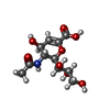  Type: D-saccharide / Mass: 291.255 Da / Num. of mol.: 4 / Source method: obtained synthetically / Formula: C11H17NO8 / Feature type: SUBJECT OF INVESTIGATION Type: D-saccharide / Mass: 291.255 Da / Num. of mol.: 4 / Source method: obtained synthetically / Formula: C11H17NO8 / Feature type: SUBJECT OF INVESTIGATION#3: Water | ChemComp-HOH / |  Mass: 18.015 Da / Num. of mol.: 1040 / Source method: isolated from a natural source / Formula: H2O Mass: 18.015 Da / Num. of mol.: 1040 / Source method: isolated from a natural source / Formula: H2OHas ligand of interest | Y | Has protein modification | N | |
|---|
-Experimental details
-Experiment
| Experiment | Method: X-RAY DIFFRACTION / Number of used crystals: 1 |
|---|
- Sample preparation
| Crystal | Density Matthews: 2.28 Å3/Da / Density % sol: 46 % / Description: P 1 21 1 space group |
|---|---|
| Crystal grow | Temperature: 289.15 K / Method: vapor diffusion, sitting drop / pH: 4 Details: After 6 weeks, crystals formed on the PEGIon tray condition 0.2M sodium malonate pH 4.0, 20% w/v Polyethylene glycol 3,350 at 289.15K |
-Data collection
| Diffraction | Mean temperature: 277.15 K / Serial crystal experiment: N |
|---|---|
| Diffraction source | Source: SYNCHROTRON / Site: APS  / Beamline: 23-ID-D / Wavelength: 0.99 Å / Beamline: 23-ID-D / Wavelength: 0.99 Å |
| Detector | Type: DECTRIS PILATUS 6M / Detector: PIXEL / Date: Dec 6, 2019 |
| Radiation | Protocol: SINGLE WAVELENGTH / Monochromatic (M) / Laue (L): M / Scattering type: x-ray |
| Radiation wavelength | Wavelength: 0.99 Å / Relative weight: 1 |
| Reflection | Resolution: 2.1→78.51 Å / Num. obs: 126161 / % possible obs: 99.76 % / Redundancy: 4.1 % / Biso Wilson estimate: 35.51 Å2 / CC1/2: 0.987 / Rmerge(I) obs: 0.113 / Rpim(I) all: 0.101 / Rrim(I) all: 0.152 / Χ2: 0.47 / Net I/σ(I): 1.4 |
| Reflection shell | Resolution: 2.1→7.25 Å / Rmerge(I) obs: 0.113 / Mean I/σ(I) obs: 7.3 / Num. unique obs: 5927 / CC1/2: 0.987 / Rpim(I) all: 0.101 / Rrim(I) all: 0.152 / Χ2: 1.07 / % possible all: 96.7 |
- Processing
| Software |
| |||||||||||||||||||||||||||||||||||||||||||||||||||||||||||||||||||||||||||||||||||||||||||||||||||||||||||||||||||||||||||||||||||||||||||||||||||||||||||||||||||||||||||||||||||||||||||||||||||||||||||||||||||||||||
|---|---|---|---|---|---|---|---|---|---|---|---|---|---|---|---|---|---|---|---|---|---|---|---|---|---|---|---|---|---|---|---|---|---|---|---|---|---|---|---|---|---|---|---|---|---|---|---|---|---|---|---|---|---|---|---|---|---|---|---|---|---|---|---|---|---|---|---|---|---|---|---|---|---|---|---|---|---|---|---|---|---|---|---|---|---|---|---|---|---|---|---|---|---|---|---|---|---|---|---|---|---|---|---|---|---|---|---|---|---|---|---|---|---|---|---|---|---|---|---|---|---|---|---|---|---|---|---|---|---|---|---|---|---|---|---|---|---|---|---|---|---|---|---|---|---|---|---|---|---|---|---|---|---|---|---|---|---|---|---|---|---|---|---|---|---|---|---|---|---|---|---|---|---|---|---|---|---|---|---|---|---|---|---|---|---|---|---|---|---|---|---|---|---|---|---|---|---|---|---|---|---|---|---|---|---|---|---|---|---|---|---|---|---|---|---|---|---|---|
| Refinement | Method to determine structure: MOLECULAR REPLACEMENT / Resolution: 2.1→78.51 Å / SU ML: 0.3201 / Cross valid method: FREE R-VALUE / σ(F): 1.33 / Phase error: 26.9709 Stereochemistry target values: GeoStd + Monomer Library + CDL v1.2
| |||||||||||||||||||||||||||||||||||||||||||||||||||||||||||||||||||||||||||||||||||||||||||||||||||||||||||||||||||||||||||||||||||||||||||||||||||||||||||||||||||||||||||||||||||||||||||||||||||||||||||||||||||||||||
| Solvent computation | Shrinkage radii: 0.9 Å / VDW probe radii: 1.11 Å / Solvent model: FLAT BULK SOLVENT MODEL | |||||||||||||||||||||||||||||||||||||||||||||||||||||||||||||||||||||||||||||||||||||||||||||||||||||||||||||||||||||||||||||||||||||||||||||||||||||||||||||||||||||||||||||||||||||||||||||||||||||||||||||||||||||||||
| Displacement parameters | Biso mean: 40.71 Å2 | |||||||||||||||||||||||||||||||||||||||||||||||||||||||||||||||||||||||||||||||||||||||||||||||||||||||||||||||||||||||||||||||||||||||||||||||||||||||||||||||||||||||||||||||||||||||||||||||||||||||||||||||||||||||||
| Refinement step | Cycle: LAST / Resolution: 2.1→78.51 Å
| |||||||||||||||||||||||||||||||||||||||||||||||||||||||||||||||||||||||||||||||||||||||||||||||||||||||||||||||||||||||||||||||||||||||||||||||||||||||||||||||||||||||||||||||||||||||||||||||||||||||||||||||||||||||||
| Refine LS restraints |
| |||||||||||||||||||||||||||||||||||||||||||||||||||||||||||||||||||||||||||||||||||||||||||||||||||||||||||||||||||||||||||||||||||||||||||||||||||||||||||||||||||||||||||||||||||||||||||||||||||||||||||||||||||||||||
| LS refinement shell |
|
