 Movie
Movie Controller

[English] 日本語
 Yorodumi
- PDB-9dal: Human norovirus GII.3 R112A mutant protease in complex with rupin... -
+ Open data

- Basic information
| Entry | Database: PDB / ID: 9dal | |||||||||
|---|---|---|---|---|---|---|---|---|---|---|
| Title | Human norovirus GII.3 R112A mutant protease in complex with rupintrivir | |||||||||
 Components Components | Peptidase C37 | |||||||||
 Keywords Keywords | VIRAL PROTEIN / Viral protease with inhibitor | |||||||||
| Function / homology |  Function and homology information Function and homology informationribonucleoside triphosphate phosphatase activity / RNA helicase activity / cysteine-type endopeptidase activity / viral RNA genome replication / RNA-directed RNA polymerase activity / DNA-templated transcription / proteolysis / RNA binding / ATP binding Similarity search - Function | |||||||||
| Biological species |  Norovirus Norovirus | |||||||||
| Method |  X-RAY DIFFRACTION / SYNCHROTRON / MOLECULAR REPLACEMENT / Resolution: 2.6 Å X-RAY DIFFRACTION / SYNCHROTRON / MOLECULAR REPLACEMENT / Resolution: 2.6 Å | |||||||||
 Authors Authors | Pham, S.H. / Neetu, N. / Sankaran, B. / Prasad, B.V.V. | |||||||||
| Funding support |  United States, 2items United States, 2items
| |||||||||
 Citation Citation | Journal: J.Virol. / Year: 2025 Title: Conformational flexibility is a critical factor in designing broad-spectrum human norovirus protease inhibitors. Authors: Pham, S. / Zhao, B. / Neetu, N. / Sankaran, B. / Patil, K. / Ramani, S. / Song, Y. / Estes, M.K. / Palzkill, T. / Prasad, B.V.V. | |||||||||
| History |
|
- Structure visualization
| Structure viewer | Molecule: MolmilJmol/JSmol |
|---|
- Downloads & links
-Download
| PDBx/mmCIF format | 9dal.cif.gz | 112.1 KB | Display | PDBx/mmCIF format |
|---|---|---|---|---|
| PDB format | pdb9dal.ent.gz | 83.7 KB | Display | PDB format |
| PDBx/mmJSON format | 9dal.json.gz | Tree view | PDBx/mmJSON format | |
| Others |  Other downloads Other downloads |
-Validation report
| Arichive directory | https://data.pdbj.org/pub/pdb/validation_reports/da/9dalftp://data.pdbj.org/pub/pdb/validation_reports/da/9dal | HTTPS FTP |
|---|
-Related structure data
| Related structure data | 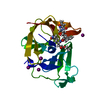 9d9tC  9d9yC  9da0C  9da7C  9dajC  9dapC 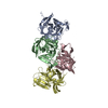 9deyC  9df5C C: citing same article ( |
|---|---|
| Similar structure data |
-Links
 PDBj
PDBj
- Assembly
| Deposited unit | 
| ||||||||||||
|---|---|---|---|---|---|---|---|---|---|---|---|---|---|
| 1 |
| ||||||||||||
| Unit cell |
|
-Components
| #1: Protein | Mass: 19415.340 Da / Num. of mol.: 1 / Mutation: R112A Source method: isolated from a genetically manipulated source Source: (gene. exp.) Norovirus / Production host:  |
|---|---|
| #2: Chemical | ChemComp-AG7 /   Mass: 600.678 Da / Num. of mol.: 1 / Source method: obtained synthetically / Formula: C31H41FN4O7 / Feature type: SUBJECT OF INVESTIGATION / Comment: antivirus, protease inhibitor*YM Mass: 600.678 Da / Num. of mol.: 1 / Source method: obtained synthetically / Formula: C31H41FN4O7 / Feature type: SUBJECT OF INVESTIGATION / Comment: antivirus, protease inhibitor*YM |
| Has ligand of interest | Y |
| Has protein modification | N |
-Experimental details
-Experiment
| Experiment | Method: X-RAY DIFFRACTION / Number of used crystals: 1 |
|---|
- Sample preparation
| Crystal | Density Matthews: 3.1 Å3/Da / Density % sol: 60.34 % |
|---|---|
| Crystal grow | Temperature: 298 K / Method: vapor diffusion, hanging drop / pH: 9 Details: 0.1 M BICINE pH 9.0, 10% (w/v) PEG 20000; 2%(v/v) 1,4-Dioxane |
-Data collection
| Diffraction | Mean temperature: 100 K / Serial crystal experiment: N |
|---|---|
| Diffraction source | Source: SYNCHROTRON / Site: ALS / Beamline: 5.0.1 / Wavelength: 0.97741 Å |
| Detector | Type: DECTRIS PILATUS3 2M / Detector: PIXEL / Date: May 18, 2023 |
| Radiation | Protocol: SINGLE WAVELENGTH / Monochromatic (M) / Laue (L): M / Scattering type: x-ray |
| Radiation wavelength | Wavelength: 0.97741 Å / Relative weight: 1 |
| Reflection | Resolution: 2.6→41.16 Å / Num. obs: 7621 / % possible obs: 100 % / Redundancy: 18 % / Biso Wilson estimate: 71.56 Å2 / CC1/2: 1 / CC star: 1 / Rmerge(I) obs: 0.079 / Rpim(I) all: 0.019 / Rrim(I) all: 0.081 / Net I/σ(I): 26.5 |
| Reflection shell | Resolution: 2.6→2.64 Å / Mean I/σ(I) obs: 1.8 / Num. unique obs: 378 / CC1/2: 0.352 / CC star: 0.722 / % possible all: 100 |
- Processing
| Software |
| ||||||||||||||||||||||||||||||||||||||||||
|---|---|---|---|---|---|---|---|---|---|---|---|---|---|---|---|---|---|---|---|---|---|---|---|---|---|---|---|---|---|---|---|---|---|---|---|---|---|---|---|---|---|---|---|
| Refinement | Method to determine structure: MOLECULAR REPLACEMENT / Resolution: 2.6→41.16 Å / SU ML: 0.3203 / Cross valid method: FREE R-VALUE / σ(F): 1.34 / Phase error: 29.8946 Stereochemistry target values: GeoStd + Monomer Library + CDL v1.2
| ||||||||||||||||||||||||||||||||||||||||||
| Solvent computation | Shrinkage radii: 0.9 Å / VDW probe radii: 1.1 Å / Solvent model: FLAT BULK SOLVENT MODEL | ||||||||||||||||||||||||||||||||||||||||||
| Displacement parameters | Biso mean: 77.35 Å2 | ||||||||||||||||||||||||||||||||||||||||||
| Refinement step | Cycle: LAST / Resolution: 2.6→41.16 Å
| ||||||||||||||||||||||||||||||||||||||||||
| Refine LS restraints |
| ||||||||||||||||||||||||||||||||||||||||||
| LS refinement shell |
|
