 Movie
Movie Controller


+ Open data

- Basic information
| Entry | Database: PDB / ID: 9da1 | |||||||||
|---|---|---|---|---|---|---|---|---|---|---|
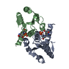
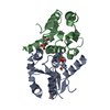
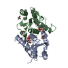
| Title | Crystal structure of human DNPH1 bound to inhibitor 1a | |||||||||
 Components Components | 5-hydroxymethyl-dUMP N-hydrolase | |||||||||
 Keywords Keywords | HYDROLASE/INHIBITOR / Inhibitor / Complex / HYDROLASE / HYDROLASE-INHIBITOR complex | |||||||||
| Function / homology |  Function and homology information Function and homology informationpurine nucleotide catabolic process / deoxyribonucleoside monophosphate catabolic process / 5-hydroxymethyl-dUMP N-hydrolase activity / nucleoside salvage / dGMP catabolic process / Purine catabolism / allantoin metabolic process / Hydrolases; Glycosylases; Hydrolysing N-glycosyl compounds / epithelial cell differentiation / positive regulation of cell growth ...purine nucleotide catabolic process / deoxyribonucleoside monophosphate catabolic process / 5-hydroxymethyl-dUMP N-hydrolase activity / nucleoside salvage / dGMP catabolic process / Purine catabolism / allantoin metabolic process / Hydrolases; Glycosylases; Hydrolysing N-glycosyl compounds / epithelial cell differentiation / positive regulation of cell growth / protein homodimerization activity / extracellular exosome / identical protein binding / nucleus / cytoplasm / cytosol Similarity search - Function | |||||||||
| Biological species |  Homo sapiens (human) Homo sapiens (human) | |||||||||
| Method |  X-RAY DIFFRACTION / SYNCHROTRON / MOLECULAR REPLACEMENT / Resolution: 1.47 Å X-RAY DIFFRACTION / SYNCHROTRON / MOLECULAR REPLACEMENT / Resolution: 1.47 Å | |||||||||
 Authors Authors | Wagner, A.G. / Schramm, V.L. / Almo, S.C. / Ghosh, A. | |||||||||
| Funding support |  United States, 2items United States, 2items
| |||||||||
 Citation Citation | Journal: J.Med.Chem. / Year: 2025 Title: Transition State Analogs of Human DNPH1 Reveal Two Electrophile Migration Mechanisms. Authors: Wagner, A.G. / Lang, T.B.D. / Ledingham, E.T. / Ghosh, A. / Brooks, D. / Eskandari, R. / Suthagar, K. / Almo, S.C. / Lamiable-Oulaidi, F. / Tyler, P.C. / Schramm, V.L. | |||||||||
| History |
|
- Structure visualization
| Structure viewer | Molecule: MolmilJmol/JSmol |
|---|
- Downloads & links
-Download
| PDBx/mmCIF format | 9da1.cif.gz | 168 KB | Display | PDBx/mmCIF format |
|---|---|---|---|---|
| PDB format | pdb9da1.ent.gz | Display | PDB format | |
| PDBx/mmJSON format | 9da1.json.gz | Tree view | PDBx/mmJSON format | |
| Others |  Other downloads Other downloads |
-Validation report
| Summary document | 9da1_validation.pdf.gz | 1.1 MB | Display | wwPDB validaton report |
|---|---|---|---|---|
| Full document | 9da1_full_validation.pdf.gz | 1.1 MB | Display | |
| Data in XML | 9da1_validation.xml.gz | 16.4 KB | Display | |
| Data in CIF | 9da1_validation.cif.gz | 22.1 KB | Display | |
| Arichive directory | https://data.pdbj.org/pub/pdb/validation_reports/da/9da1ftp://data.pdbj.org/pub/pdb/validation_reports/da/9da1 | HTTPS FTP |
-Related structure data


-Links
 PDBj
PDBj- Assembly
| Deposited unit | 
| ||||||||||||
|---|---|---|---|---|---|---|---|---|---|---|---|---|---|
| 1 |
| ||||||||||||
| Unit cell |
| ||||||||||||
| Components on special symmetry positions |
|
-Components
| #1: Protein | Mass: 16224.247 Da / Num. of mol.: 2 Source method: isolated from a genetically manipulated source Source: (gene. exp.) Homo sapiens (human) / Gene: DNPH1, C6orf108, RCL / Plasmid: pET-28a(+) / Details (production host): LIC / Production host:  References: UniProt: O43598, Hydrolases; Glycosylases; Hydrolysing N-glycosyl compounds #2: Chemical | Mass: 354.207 Da / Num. of mol.: 2 / Source method: obtained synthetically / Formula: C10H15N2O10P / Feature type: SUBJECT OF INVESTIGATION #3: Water | ChemComp-HOH / |  Mass: 18.015 Da / Num. of mol.: 217 / Source method: isolated from a natural source / Formula: H2O Mass: 18.015 Da / Num. of mol.: 217 / Source method: isolated from a natural source / Formula: H2OHas ligand of interest | Y | Has protein modification | N | |
|---|
-Experimental details
-Experiment
| Experiment | Method: X-RAY DIFFRACTION / Number of used crystals: 1 |
|---|
- Sample preparation
| Crystal | Density Matthews: 1.95 Å3/Da / Density % sol: 36.91 % |
|---|---|
| Crystal grow | Temperature: 292 K / Method: vapor diffusion, sitting drop / pH: 4.5 Details: 25.1% (w/v) PEG 3350, 2.73% (v/v) Methanol and Sodium Acetate |
-Data collection
| Diffraction | Mean temperature: 100 K / Serial crystal experiment: N |
|---|---|
| Diffraction source | Source: SYNCHROTRON / Site: NSLS-II / Beamline: 17-ID-2 / Wavelength: 0.92 Å |
| Detector | Type: DECTRIS EIGER X 16M / Detector: PIXEL / Date: Jul 17, 2023 |
| Radiation | Protocol: SINGLE WAVELENGTH / Monochromatic (M) / Laue (L): M / Scattering type: x-ray |
| Radiation wavelength | Wavelength: 0.92 Å / Relative weight: 1 |
| Reflection | Resolution: 1.47→22.82 Å / Num. obs: 43023 / % possible obs: 99.7 % / Redundancy: 9.4 % / CC1/2: 0.998 / Rmerge(I) obs: 0.083 / Rpim(I) all: 0.028 / Rrim(I) all: 0.088 / Χ2: 0.99 / Net I/σ(I): 14.8 / Num. measured all: 404482 |
| Reflection shell | Resolution: 1.47→1.5 Å / % possible obs: 94.4 % / Redundancy: 9.4 % / Rmerge(I) obs: 0.776 / Num. measured all: 18516 / Num. unique obs: 1960 / CC1/2: 0.846 / Rpim(I) all: 0.261 / Rrim(I) all: 0.82 / Χ2: 1.11 / Net I/σ(I) obs: 3.2 |
- Processing
| Software |
| |||||||||||||||||||||||||||||||||||||||||||||||||||||||||||||||||||||||||||||||||||||||||||||||||||||||||||||||||||||||||||||||||||||||||||||||||||||||||||||||||||||||||||||||||||||||||||||||||||||||||||||||||||||||||||||||||||||||||||||||||||||||||||||||||||||||||||||||||||||||||||||||||||||||||||||||||||||||||||||||||||||
|---|---|---|---|---|---|---|---|---|---|---|---|---|---|---|---|---|---|---|---|---|---|---|---|---|---|---|---|---|---|---|---|---|---|---|---|---|---|---|---|---|---|---|---|---|---|---|---|---|---|---|---|---|---|---|---|---|---|---|---|---|---|---|---|---|---|---|---|---|---|---|---|---|---|---|---|---|---|---|---|---|---|---|---|---|---|---|---|---|---|---|---|---|---|---|---|---|---|---|---|---|---|---|---|---|---|---|---|---|---|---|---|---|---|---|---|---|---|---|---|---|---|---|---|---|---|---|---|---|---|---|---|---|---|---|---|---|---|---|---|---|---|---|---|---|---|---|---|---|---|---|---|---|---|---|---|---|---|---|---|---|---|---|---|---|---|---|---|---|---|---|---|---|---|---|---|---|---|---|---|---|---|---|---|---|---|---|---|---|---|---|---|---|---|---|---|---|---|---|---|---|---|---|---|---|---|---|---|---|---|---|---|---|---|---|---|---|---|---|---|---|---|---|---|---|---|---|---|---|---|---|---|---|---|---|---|---|---|---|---|---|---|---|---|---|---|---|---|---|---|---|---|---|---|---|---|---|---|---|---|---|---|---|---|---|---|---|---|---|---|---|---|---|---|---|---|---|---|---|---|---|---|---|---|---|---|---|---|---|---|---|---|---|---|---|---|---|---|---|---|---|---|---|---|---|---|---|---|---|---|---|---|---|---|---|---|---|---|---|---|---|---|---|---|---|---|---|
| Refinement | Method to determine structure: MOLECULAR REPLACEMENT / Resolution: 1.47→22.82 Å / SU ML: 0.1 / Cross valid method: FREE R-VALUE / σ(F): 1.34 / Phase error: 15.67 / Stereochemistry target values: ML
| |||||||||||||||||||||||||||||||||||||||||||||||||||||||||||||||||||||||||||||||||||||||||||||||||||||||||||||||||||||||||||||||||||||||||||||||||||||||||||||||||||||||||||||||||||||||||||||||||||||||||||||||||||||||||||||||||||||||||||||||||||||||||||||||||||||||||||||||||||||||||||||||||||||||||||||||||||||||||||||||||||||
| Solvent computation | Shrinkage radii: 0.9 Å / VDW probe radii: 1.1 Å / Solvent model: FLAT BULK SOLVENT MODEL | |||||||||||||||||||||||||||||||||||||||||||||||||||||||||||||||||||||||||||||||||||||||||||||||||||||||||||||||||||||||||||||||||||||||||||||||||||||||||||||||||||||||||||||||||||||||||||||||||||||||||||||||||||||||||||||||||||||||||||||||||||||||||||||||||||||||||||||||||||||||||||||||||||||||||||||||||||||||||||||||||||||
| Refinement step | Cycle: LAST / Resolution: 1.47→22.82 Å
| |||||||||||||||||||||||||||||||||||||||||||||||||||||||||||||||||||||||||||||||||||||||||||||||||||||||||||||||||||||||||||||||||||||||||||||||||||||||||||||||||||||||||||||||||||||||||||||||||||||||||||||||||||||||||||||||||||||||||||||||||||||||||||||||||||||||||||||||||||||||||||||||||||||||||||||||||||||||||||||||||||||
| Refine LS restraints |
| |||||||||||||||||||||||||||||||||||||||||||||||||||||||||||||||||||||||||||||||||||||||||||||||||||||||||||||||||||||||||||||||||||||||||||||||||||||||||||||||||||||||||||||||||||||||||||||||||||||||||||||||||||||||||||||||||||||||||||||||||||||||||||||||||||||||||||||||||||||||||||||||||||||||||||||||||||||||||||||||||||||
| LS refinement shell |
| |||||||||||||||||||||||||||||||||||||||||||||||||||||||||||||||||||||||||||||||||||||||||||||||||||||||||||||||||||||||||||||||||||||||||||||||||||||||||||||||||||||||||||||||||||||||||||||||||||||||||||||||||||||||||||||||||||||||||||||||||||||||||||||||||||||||||||||||||||||||||||||||||||||||||||||||||||||||||||||||||||||
| Refinement TLS params. | Method: refined / Refine-ID: X-RAY DIFFRACTION
| |||||||||||||||||||||||||||||||||||||||||||||||||||||||||||||||||||||||||||||||||||||||||||||||||||||||||||||||||||||||||||||||||||||||||||||||||||||||||||||||||||||||||||||||||||||||||||||||||||||||||||||||||||||||||||||||||||||||||||||||||||||||||||||||||||||||||||||||||||||||||||||||||||||||||||||||||||||||||||||||||||||
| Refinement TLS group |
|
