 Movie
Movie Controller

[English] 日本語
 Yorodumi
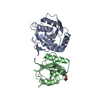
- PDB-9d91: Crystal structure of L-asparaginase from Streptococcus pneumoniae... -
+ Open data

- Basic information
| Entry | Database: PDB / ID: 9d91 | ||||||
|---|---|---|---|---|---|---|---|
| Title | Crystal structure of L-asparaginase from Streptococcus pneumoniae TIGR4 | ||||||
 Components Components | Asparaginase | ||||||
 Keywords Keywords | HYDROLASE / Structural Genomics / Center for Structural Biology of Infectious Diseases / CSBID | ||||||
| Function / homology |  Function and homology information Function and homology information | ||||||
| Biological species |   Streptococcus pneumoniae (bacteria) Streptococcus pneumoniae (bacteria) | ||||||
| Method |  X-RAY DIFFRACTION / SYNCHROTRON / MOLECULAR REPLACEMENT / Resolution: 1.62 Å X-RAY DIFFRACTION / SYNCHROTRON / MOLECULAR REPLACEMENT / Resolution: 1.62 Å | ||||||
 Authors Authors | Gade, P. / Endres, M. / Babnigg, G. / Joachimiak, A. / Center for Structural Biology of Infectious Diseases (CSBID) | ||||||
| Funding support |  United States, 1items United States, 1items
| ||||||
 Citation Citation | Journal: To Be Published Title: Crystal structure of L-asparaginase from Streptococcus pneumoniae TIGR4 Authors: Gade, P. / Endres, M. / Babnigg, G. / Joachimiak, A. | ||||||
| History |
|
- Structure visualization
| Structure viewer | Molecule: MolmilJmol/JSmol |
|---|
- Downloads & links
-Download
| PDBx/mmCIF format | 9d91.cif.gz | 166.9 KB | Display | PDBx/mmCIF format |
|---|---|---|---|---|
| PDB format | pdb9d91.ent.gz | 108.9 KB | Display | PDB format |
| PDBx/mmJSON format | 9d91.json.gz | Tree view | PDBx/mmJSON format | |
| Others |  Other downloads Other downloads |
-Validation report
| Summary document | 9d91_validation.pdf.gz | 432.6 KB | Display | wwPDB validaton report |
|---|---|---|---|---|
| Full document | 9d91_full_validation.pdf.gz | 432.6 KB | Display | |
| Data in XML | 9d91_validation.xml.gz | 17.1 KB | Display | |
| Data in CIF | 9d91_validation.cif.gz | 24.1 KB | Display | |
| Arichive directory | https://data.pdbj.org/pub/pdb/validation_reports/d9/9d91ftp://data.pdbj.org/pub/pdb/validation_reports/d9/9d91 | HTTPS FTP |
-Related structure data
| Related structure data | |
|---|---|
| Similar structure data |

-Links
 PDBj
PDBj- Assembly
| Deposited unit | 
| ||||||||||||
|---|---|---|---|---|---|---|---|---|---|---|---|---|---|
| 1 |
| ||||||||||||
| Unit cell |
| ||||||||||||
| Components on special symmetry positions |
|
-Components
| #1: Protein | Mass: 34907.055 Da / Num. of mol.: 1 Source method: isolated from a genetically manipulated source Source: (gene. exp.) Streptococcus pneumoniae (bacteria)Gene: ansB, A5N45_01685, AZJ70_05345, AZK02_05425, ERS019316_00818, ERS019420_01259, ERS021218_01180, GM536_02705, GM543_02955, GM545_09810, RLD18_05820, SAMEA3171064_00528, SAMEA3353485_02125, ...Gene: ansB, A5N45_01685, AZJ70_05345, AZK02_05425, ERS019316_00818, ERS019420_01259, ERS021218_01180, GM536_02705, GM543_02955, GM545_09810, RLD18_05820, SAMEA3171064_00528, SAMEA3353485_02125, SAMEA3353631_01742, SAMEA3354366_01577, SAMEA3389353_01902, SAMEA4038883_00005 Production host: | ||||||||
|---|---|---|---|---|---|---|---|---|---|
| #2: Chemical |   Mass: 62.068 Da / Num. of mol.: 2 / Source method: obtained synthetically / Formula: C2H6O2 Mass: 62.068 Da / Num. of mol.: 2 / Source method: obtained synthetically / Formula: C2H6O2#3: Chemical | ChemComp-SO4 /   Mass: 96.063 Da / Num. of mol.: 4 / Source method: obtained synthetically / Formula: SO4 Mass: 96.063 Da / Num. of mol.: 4 / Source method: obtained synthetically / Formula: SO4#4: Water | ChemComp-HOH / |  Mass: 18.015 Da / Num. of mol.: 226 / Source method: isolated from a natural source / Formula: H2O Mass: 18.015 Da / Num. of mol.: 226 / Source method: isolated from a natural source / Formula: H2OHas ligand of interest | N | Has protein modification | N | |
-Experimental details
-Experiment
| Experiment | Method: X-RAY DIFFRACTION / Number of used crystals: 1 |
|---|
- Sample preparation
| Crystal | Density Matthews: 1.91 Å3/Da / Density % sol: 35.66 % |
|---|---|
| Crystal grow | Temperature: 289 K / Method: vapor diffusion, sitting drop / pH: 7 Details: 0.2 M Lithium Sulfate, 0.1 M Tris HCl pH 7.0, 2 M Ammonium Sulfate |
-Data collection
| Diffraction | Mean temperature: 100 K / Serial crystal experiment: N |
|---|---|
| Diffraction source | Source: SYNCHROTRON / Site: NSLS-II / Beamline: 19-ID / Wavelength: 0.97934 Å |
| Detector | Type: DECTRIS EIGER2 XE 9M / Detector: PIXEL / Date: Aug 5, 2024 |
| Radiation | Protocol: SINGLE WAVELENGTH / Monochromatic (M) / Laue (L): M / Scattering type: x-ray |
| Radiation wavelength | Wavelength: 0.97934 Å / Relative weight: 1 |
| Reflection | Resolution: 1.62→50 Å / Num. obs: 34185 / % possible obs: 99.8 % / Redundancy: 6.1 % / Biso Wilson estimate: 13.21 Å2 / CC1/2: 0.993 / CC star: 0.998 / Rmerge(I) obs: 0.09 / Rpim(I) all: 0.042 / Rrim(I) all: 0.099 / Χ2: 0.923 / Net I/σ(I): 15.637 |
| Reflection shell | Resolution: 1.62→1.65 Å / Redundancy: 5.7 % / Rmerge(I) obs: 0.618 / Mean I/σ(I) obs: 2.8 / Num. unique obs: 34185 / CC1/2: 0.856 / CC star: 0.96 / Rpim(I) all: 0.262 / Rrim(I) all: 0.66 / % possible all: 98.8 |
- Processing
| Software |
| |||||||||||||||||||||||||||||||||||||||||||||||||||||||||||||||||||||||||||||||||||||||||||
|---|---|---|---|---|---|---|---|---|---|---|---|---|---|---|---|---|---|---|---|---|---|---|---|---|---|---|---|---|---|---|---|---|---|---|---|---|---|---|---|---|---|---|---|---|---|---|---|---|---|---|---|---|---|---|---|---|---|---|---|---|---|---|---|---|---|---|---|---|---|---|---|---|---|---|---|---|---|---|---|---|---|---|---|---|---|---|---|---|---|---|---|---|
| Refinement | Method to determine structure: MOLECULAR REPLACEMENT / Resolution: 1.62→43.82 Å / SU ML: 0.158 / Cross valid method: FREE R-VALUE / σ(F): 1.37 / Phase error: 16.6294 Stereochemistry target values: GeoStd + Monomer Library + CDL v1.2
| |||||||||||||||||||||||||||||||||||||||||||||||||||||||||||||||||||||||||||||||||||||||||||
| Solvent computation | Shrinkage radii: 0.9 Å / VDW probe radii: 1.1 Å / Solvent model: FLAT BULK SOLVENT MODEL | |||||||||||||||||||||||||||||||||||||||||||||||||||||||||||||||||||||||||||||||||||||||||||
| Displacement parameters | Biso mean: 16.88 Å2 | |||||||||||||||||||||||||||||||||||||||||||||||||||||||||||||||||||||||||||||||||||||||||||
| Refinement step | Cycle: LAST / Resolution: 1.62→43.82 Å
| |||||||||||||||||||||||||||||||||||||||||||||||||||||||||||||||||||||||||||||||||||||||||||
| Refine LS restraints |
| |||||||||||||||||||||||||||||||||||||||||||||||||||||||||||||||||||||||||||||||||||||||||||
| LS refinement shell |
| |||||||||||||||||||||||||||||||||||||||||||||||||||||||||||||||||||||||||||||||||||||||||||
| Refinement TLS params. | Method: refined / Origin x: -16.1971134209 Å / Origin y: -27.9061777086 Å / Origin z: -16.5508964298 Å
| |||||||||||||||||||||||||||||||||||||||||||||||||||||||||||||||||||||||||||||||||||||||||||
| Refinement TLS group | Selection details: all |
