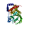
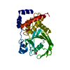
Entry Database : PDB / ID : 9cyrTitle Crystal structure of D245G mutant human PTP1B (PTPN1) at room temperature (298 K) Tyrosine-protein phosphatase non-receptor type 1 Keywords / / / / Function / homology Function Domain/homology Component
/ / / / / / / / / / / / / / / / / / / / / / / / / / / / / / / / / / / / / / / / / / / / / / / / / / / / / / / / / / / / / / / / / / / / / / / / / / / / / / / / / / / / / / / / / / / / / / / / / / / / / / / / / / Biological species Homo sapiens (human)Method / / / Resolution : 1.65 Å Authors Ebrahim, A. / Perdikari, A. / Woods, V.A. / Lawler, K. / Bounds, R. / Shah, D.S. / Singh, N.I. / Mehlman, T. / Riley, B.T. / Sharma, S. ...Ebrahim, A. / Perdikari, A. / Woods, V.A. / Lawler, K. / Bounds, R. / Shah, D.S. / Singh, N.I. / Mehlman, T. / Riley, B.T. / Sharma, S. / Morris, J.W. / Keogh, J.M. / Henning, E. / Smith, M. / Farooqi, I.S. / Keedy, D.A. Funding support Organization Grant number Country National Institutes of Health/National Institute of General Medical Sciences (NIH/NIGMS) GM133769 Wellcome Trust 207462/Z/17/Z
Journal : J.Biol.Chem. / Year : 2025Title : Structures of human protein tyrosine phosphatase variants reveal targetable allosteric sites.
Authors: Perdikari, A. / Woods, V.A. / Ebrahim, A. / Lawler, K. / Bounds, R. / Shah, D.S. / Singh, N.I. / Mehlman, T.S. / Riley, B.T. / Sharma, S. / Morris, J.W. / Keogh, J.M. / Henning, E. / Smith, ... Authors : Perdikari, A. / Woods, V.A. / Ebrahim, A. / Lawler, K. / Bounds, R. / Shah, D.S. / Singh, N.I. / Mehlman, T.S. / Riley, B.T. / Sharma, S. / Morris, J.W. / Keogh, J.M. / Henning, E. / Smith, M. / Farooqi, I.S. / Keedy, D.A. #1: Journal : Biorxiv / Year : 2025Title : Structures of human PTP1B variants reveal allosteric sites to target for weight loss therapy.
Authors :
Perdikari, A. / Woods, V.A. / Ebrahim, A. / Lawler, K. / Bounds, R. / Singh, N.I. / Mehlman, T.S. / Riley, B.T. / Sharma, S. / Morris, J.W. / Keogh, J.M. / Henning, E. / Smith, M. / Farooqi, I.S. / Keedy, D.A. History Deposition Aug 2, 2024 Deposition site / Processing site Revision 1.0 Aug 21, 2024 Provider / Type Revision 1.1 Aug 28, 2024 Group / Category / citation_authorItem _citation.pdbx_database_id_PubMed / _citation.title ... _citation.pdbx_database_id_PubMed / _citation.title / _citation_author.identifier_ORCID / _citation_author.name Revision 1.2 Sep 4, 2024 Group / Category / Item Revision 2.0 Apr 16, 2025 Group Atomic model / Data collection ... Atomic model / Data collection / Database references / Derived calculations / Non-polymer description / Polymer sequence / Structure summary Category atom_site / chem_comp ... atom_site / chem_comp / chem_comp_atom / chem_comp_bond / citation / citation_author / entity / entity_poly / entity_poly_seq / pdbx_entity_nonpoly / pdbx_entry_details / pdbx_modification_feature / pdbx_nonpoly_scheme / pdbx_poly_seq_scheme / pdbx_struct_assembly_gen / pdbx_struct_conn_angle / struct_asym / struct_conn Item _atom_site.B_iso_or_equiv / _atom_site.Cartn_x ... _atom_site.B_iso_or_equiv / _atom_site.Cartn_x / _atom_site.Cartn_y / _atom_site.Cartn_z / _atom_site.auth_atom_id / _atom_site.auth_comp_id / _atom_site.auth_seq_id / _atom_site.group_PDB / _atom_site.label_alt_id / _atom_site.label_asym_id / _atom_site.label_atom_id / _atom_site.label_comp_id / _atom_site.label_entity_id / _atom_site.label_seq_id / _atom_site.occupancy / _atom_site.type_symbol / _chem_comp.formula / _chem_comp.formula_weight / _chem_comp.id / _chem_comp.mon_nstd_flag / _chem_comp.name / _chem_comp.type / _citation.year / _citation_author.identifier_ORCID / _entity_poly.nstd_monomer / _entity_poly.pdbx_seq_one_letter_code / _entity_poly_seq.mon_id / _pdbx_entry_details.has_protein_modification / _pdbx_poly_seq_scheme.mon_id / _pdbx_poly_seq_scheme.pdb_mon_id / _pdbx_struct_assembly_gen.asym_id_list / _pdbx_struct_conn_angle.ptnr1_label_asym_id / _pdbx_struct_conn_angle.ptnr2_auth_seq_id / _pdbx_struct_conn_angle.ptnr2_label_asym_id / _pdbx_struct_conn_angle.ptnr3_label_asym_id Revision 2.1 Nov 26, 2025 Group / Structure summary / Category / citation / citation_authorRevision 2.2 Dec 10, 2025 Group / Category / Item
Show all Show less
 Movie
Movie Controller

 Yorodumi
 Open data
 Basic information
 Components
 Keywords
 Function and homology information
 Homo sapiens (human)
 X-RAY DIFFRACTION /
  Authors
 United States,
  United Kingdom, 2items
  Citation
 Structure visualization
 Downloads & links
 Other downloads
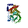
 PDBj
PDBj


 Assembly
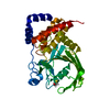


 Mass: 24.305 Da / Num. of mol.: 1 / Source method: obtained synthetically / Formula: Mg
 Mass: 18.015 Da / Num. of mol.: 140 / Source method: isolated from a natural source / Formula: H2O
 Sample preparation
 Processing