 Movie
Movie Controller

[English] 日本語
 Yorodumi
- PDB-9ctx: X-ray crystal structure of multi-drug resistant HIV-1 protease (P... -
+ Open data

- Basic information
| Entry | Database: PDB / ID: 9ctx | ||||||
|---|---|---|---|---|---|---|---|
| Title | X-ray crystal structure of multi-drug resistant HIV-1 protease (P51) in complex with Darunavir | ||||||
 Components Components | Protease | ||||||
 Keywords Keywords | VIRAL PROTEIN / HIV-1 Protease | ||||||
| Function / homology |  Function and homology information Function and homology informationhost multivesicular body / aspartic-type endopeptidase activity / virion membrane / proteolysis Similarity search - Function | ||||||
| Biological species |   Human immunodeficiency virus 1 Human immunodeficiency virus 1 | ||||||
| Method |  X-RAY DIFFRACTION / SYNCHROTRON / MOLECULAR REPLACEMENT / Resolution: 1.55 Å X-RAY DIFFRACTION / SYNCHROTRON / MOLECULAR REPLACEMENT / Resolution: 1.55 Å | ||||||
 Authors Authors | Hayashi, H. / Yedidi, R. / Bulut, H. / Das, D. / Mitsuya, H. | ||||||
| Funding support |  United States, 1items United States, 1items
| ||||||
 Citation Citation | Journal: To Be Published Title: Secondary amino-acid substitutions contribute to the emergence of HIV protease inhibitor resistance as directly as primary amino-acid substitutions. Authors: Das, D. / Hayashi, H. / Yedidi, R.S. / Bulut, H. / Mitsuya, H. | ||||||
| History |
|
- Structure visualization
| Structure viewer | Molecule: MolmilJmol/JSmol |
|---|
- Downloads & links
-Download
| PDBx/mmCIF format | 9ctx.cif.gz | 64.5 KB | Display | PDBx/mmCIF format |
|---|---|---|---|---|
| PDB format | pdb9ctx.ent.gz | 41.1 KB | Display | PDB format |
| PDBx/mmJSON format | 9ctx.json.gz | Tree view | PDBx/mmJSON format | |
| Others |  Other downloads Other downloads |
-Validation report
| Summary document | 9ctx_validation.pdf.gz | 789.3 KB | Display | wwPDB validaton report |
|---|---|---|---|---|
| Full document | 9ctx_full_validation.pdf.gz | 792.7 KB | Display | |
| Data in XML | 9ctx_validation.xml.gz | 7.9 KB | Display | |
| Data in CIF | 9ctx_validation.cif.gz | 9.8 KB | Display | |
| Arichive directory | https://data.pdbj.org/pub/pdb/validation_reports/ct/9ctxftp://data.pdbj.org/pub/pdb/validation_reports/ct/9ctx | HTTPS FTP |
-Related structure data
| Related structure data | |
|---|---|
| Similar structure data |

-Links
 PDBj
PDBj

- Assembly
| Deposited unit | 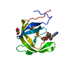
| |||||||||
|---|---|---|---|---|---|---|---|---|---|---|
| 1 | 
| |||||||||
| Unit cell |
| |||||||||
| Components on special symmetry positions |
|
-Components
| #1: Protein | Mass: 10875.717 Da / Num. of mol.: 1 Source method: isolated from a genetically manipulated source Source: (gene. exp.) Human immunodeficiency virus 1 / Gene: pol / Production host:  |
|---|---|
| #2: Chemical | ChemComp-017 / (  Mass: 547.664 Da / Num. of mol.: 1 / Source method: obtained synthetically / Formula: C27H37N3O7S / Feature type: SUBJECT OF INVESTIGATION / Comment: medication*YM Mass: 547.664 Da / Num. of mol.: 1 / Source method: obtained synthetically / Formula: C27H37N3O7S / Feature type: SUBJECT OF INVESTIGATION / Comment: medication*YM |
| #3: Chemical | ChemComp-GOL /   Mass: 92.094 Da / Num. of mol.: 1 / Source method: obtained synthetically / Formula: C3H8O3 Mass: 92.094 Da / Num. of mol.: 1 / Source method: obtained synthetically / Formula: C3H8O3 |
| #4: Water | ChemComp-HOH /  Mass: 18.015 Da / Num. of mol.: 58 / Source method: isolated from a natural source / Formula: H2O Mass: 18.015 Da / Num. of mol.: 58 / Source method: isolated from a natural source / Formula: H2O |
| Has ligand of interest | Y |
-Experimental details
-Experiment
| Experiment | Method: X-RAY DIFFRACTION / Number of used crystals: 1 |
|---|
- Sample preparation
| Crystal | Density Matthews: 2.13 Å3/Da / Density % sol: 42.29 % |
|---|---|
| Crystal grow | Temperature: 298 K / Method: vapor diffusion, hanging drop Details: 0.15 M (NH3)2SO4, 0.1 M HEPES pH7.0, 20 % (w/v) PEG 4000 |
-Data collection
| Diffraction | Mean temperature: 100 K / Serial crystal experiment: N |
|---|---|
| Diffraction source | Source: SYNCHROTRON / Site: SPring-8  / Beamline: BL24XU / Wavelength: 1 Å / Beamline: BL24XU / Wavelength: 1 Å |
| Detector | Type: RAYONIX MX225HE / Detector: CCD / Date: Jan 10, 2013 |
| Radiation | Protocol: SINGLE WAVELENGTH / Monochromatic (M) / Laue (L): M / Scattering type: x-ray |
| Radiation wavelength | Wavelength: 1 Å / Relative weight: 1 |
| Reflection | Resolution: 1.55→45.22 Å / Num. obs: 14126 / % possible obs: 98.12 % / Redundancy: 19.5 % / CC1/2: 0.998 / Net I/σ(I): 9.9 |
| Reflection shell | Resolution: 1.55→1.58 Å / Num. unique obs: 565 / CC1/2: 0.59 |
- Processing
| Software |
| ||||||||||||||||||||
|---|---|---|---|---|---|---|---|---|---|---|---|---|---|---|---|---|---|---|---|---|---|
| Refinement | Method to determine structure: MOLECULAR REPLACEMENT / Resolution: 1.55→45.22 Å / Cor.coef. Fo:Fc: 0.969 / Cor.coef. Fo:Fc free: 0.957 / Cross valid method: THROUGHOUT
| ||||||||||||||||||||
| Displacement parameters | Biso mean: 21.327 Å2
| ||||||||||||||||||||
| Refinement step | Cycle: LAST / Resolution: 1.55→45.22 Å
| ||||||||||||||||||||
| LS refinement shell | Resolution: 1.55→1.58 Å /
|
