 ムービー
ムービー コントローラー


+ データを開く

- 基本情報
基本情報
| 登録情報 | データベース: PDB / ID: 9csl | ||||||
|---|---|---|---|---|---|---|---|
| タイトル | Structure of mutant human geranylgeranyl pyrophosphate synthase (Y246D-C247L) in complex with isopentenyl pyrophosphate | ||||||
 要素 要素 | Geranylgeranyl pyrophosphate synthase | ||||||
 キーワード キーワード | TRANSFERASE / Isopentenyl transferase / Isoprenyl synthase / Isoprenoid biosynthesis / Isoprenoids | ||||||
| 機能・相同性 |  機能・相同性情報 機能・相同性情報isoprenoid metabolic process / geranylgeranyl diphosphate biosynthetic process / geranylgeranyl diphosphate synthase / geranyl diphosphate biosynthetic process / 転移酵素; メチル基以外のアルキル基またはアリル基を移すもの; - / geranylgeranyl diphosphate synthase activity / dimethylallyltranstransferase / (2E,6E)-farnesyl diphosphate synthase / Cholesterol biosynthesis / isoprenoid biosynthetic process ...isoprenoid metabolic process / geranylgeranyl diphosphate biosynthetic process / geranylgeranyl diphosphate synthase / geranyl diphosphate biosynthetic process / 転移酵素; メチル基以外のアルキル基またはアリル基を移すもの; - / geranylgeranyl diphosphate synthase activity / dimethylallyltranstransferase / (2E,6E)-farnesyl diphosphate synthase / Cholesterol biosynthesis / isoprenoid biosynthetic process / farnesyl diphosphate biosynthetic process / dimethylallyltranstransferase activity / (2E,6E)-farnesyl diphosphate synthase activity / Activation of gene expression by SREBF (SREBP) / Z disc / perinuclear region of cytoplasm / nucleoplasm / metal ion binding / identical protein binding / cytosol / cytoplasm 類似検索 - 分子機能 | ||||||
| 生物種 |  Homo sapiens (ヒト) Homo sapiens (ヒト) | ||||||
| 手法 |  X線回折 / シンクロトロン / 分子置換 / 解像度: 2.1 Å X線回折 / シンクロトロン / 分子置換 / 解像度: 2.1 Å | ||||||
 データ登録者 データ登録者 | Ezekiel, S. / Park, J. | ||||||
| 資金援助 |  カナダ, 1件 カナダ, 1件
| ||||||
 引用 引用 | ジャーナル: Plos One / 年: 2025 タイトル: Engineering dimer mutants of human geranylgeranyl pyrophosphate synthase. 著者: Ezekiel, S.J. / Searle, M. / Park, J. | ||||||
| 履歴 |
|
- 構造の表示
構造の表示
| 構造ビューア | 分子: MolmilJmol/JSmol |
|---|
- ダウンロードとリンク
-ダウンロード
| PDBx/mmCIF形式 | 9csl.cif.gz | 148.1 KB | 表示 | PDBx/mmCIF形式 |
|---|---|---|---|---|
| PDB形式 | pdb9csl.ent.gz | 110.1 KB | 表示 | PDB形式 |
| PDBx/mmJSON形式 | 9csl.json.gz | ツリー表示 | PDBx/mmJSON形式 | |
| その他 |  その他のダウンロード その他のダウンロード |
-検証レポート
| 文書・要旨 | 9csl_validation.pdf.gz | 787.9 KB | 表示 | wwPDB検証レポート |
|---|---|---|---|---|
| 文書・詳細版 | 9csl_full_validation.pdf.gz | 793.9 KB | 表示 | |
| XML形式データ | 9csl_validation.xml.gz | 30.9 KB | 表示 | |
| CIF形式データ | 9csl_validation.cif.gz | 43 KB | 表示 | |
| アーカイブディレクトリ | https://data.pdbj.org/pub/pdb/validation_reports/cs/9cslftp://data.pdbj.org/pub/pdb/validation_reports/cs/9csl | HTTPS FTP |
-関連構造データ
| 類似構造データ |
|---|
-リンク
 PDBj
PDBj

- 集合体
集合体
| 登録構造単位 | 
| ||||||||||||
|---|---|---|---|---|---|---|---|---|---|---|---|---|---|
| 1 |
| ||||||||||||
| 単位格子 |
| ||||||||||||
| 非結晶学的対称性 (NCS) | NCSドメイン:
NCSドメイン領域: Component-ID: 1 / Ens-ID: 1 / Beg auth comp-ID: MET / Beg label comp-ID: MET / End auth comp-ID: LYS / End label comp-ID: LYS / Auth asym-ID: A / Label asym-ID: A / Auth seq-ID: 1 - 296 / Label seq-ID: 23 - 318
NCSアンサンブル: (詳細: Local NCS retraints between domains: 1 2) |
-要素
| #1: タンパク質 | 分子量: 37436.504 Da / 分子数: 2 / 変異: Y246D, C247L / 由来タイプ: 組換発現 / 由来: (組換発現) Homo sapiens (ヒト) / 遺伝子: GGPS1 / 発現宿主:  参照: UniProt: O95749, 転移酵素; メチル基以外のアルキル基またはアリル基を移すもの; -, dimethylallyltranstransferase, geranylgeranyl diphosphate synthase, (2E,6E)- ...参照: UniProt: O95749, 転移酵素; メチル基以外のアルキル基またはアリル基を移すもの; -, dimethylallyltranstransferase, geranylgeranyl diphosphate synthase, (2E,6E)-farnesyl diphosphate synthase #2: 化合物 | ChemComp-IPE / | 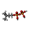  分子量: 246.092 Da / 分子数: 1 / 由来タイプ: 合成 / 式: C5H12O7P2 / タイプ: SUBJECT OF INVESTIGATION 分子量: 246.092 Da / 分子数: 1 / 由来タイプ: 合成 / 式: C5H12O7P2 / タイプ: SUBJECT OF INVESTIGATION#3: 化合物 | ChemComp-CL / |   分子量: 35.453 Da / 分子数: 1 / 由来タイプ: 合成 / 式: Cl 分子量: 35.453 Da / 分子数: 1 / 由来タイプ: 合成 / 式: Cl#4: 化合物 | ChemComp-SO4 /   分子量: 96.063 Da / 分子数: 12 / 由来タイプ: 合成 / 式: SO4 分子量: 96.063 Da / 分子数: 12 / 由来タイプ: 合成 / 式: SO4#5: 水 | ChemComp-HOH / |  分子量: 18.015 Da / 分子数: 424 / 由来タイプ: 天然 / 式: H2O 分子量: 18.015 Da / 分子数: 424 / 由来タイプ: 天然 / 式: H2O研究の焦点であるリガンドがあるか | Y | Has protein modification | Y | |
|---|
-実験情報
-実験
| 実験 | 手法: X線回折 / 使用した結晶の数: 1 |
|---|
- 試料調製
試料調製
| 結晶 | マシュー密度: 3.4 Å3/Da / 溶媒含有率: 63.84 % |
|---|---|
| 結晶化 | 温度: 295.15 K / 手法: 蒸気拡散法, シッティングドロップ法 / 詳細: 0.2 M di-ammonium phosphate, 2.2 M ammonium sulfate |
-データ収集
| 回折 | 平均測定温度: 100 K / Serial crystal experiment: N |
|---|---|
| 放射光源 | 由来: シンクロトロン / サイト: CLSI / ビームライン: 08ID-1 / 波長: 0.97949 Å |
| 検出器 | タイプ: DECTRIS PILATUS3 S 6M / 検出器: PIXEL / 日付: 2019年8月16日 |
| 放射 | プロトコル: SINGLE WAVELENGTH / 単色(M)・ラウエ(L): M / 散乱光タイプ: x-ray |
| 放射波長 | 波長: 0.97949 Å / 相対比: 1 |
| 反射 | 解像度: 2.1→95.56 Å / Num. obs: 60724 / % possible obs: 99.9 % / 冗長度: 19.6 % / Biso Wilson estimate: 40.173 Å2 / CC1/2: 1 / Rmerge(I) obs: 0.101 / Rpim(I) all: 0.023 / Rrim(I) all: 0.104 / Net I/σ(I): 20.4 |
| 反射 シェル | 解像度: 2.1→2.14 Å / 冗長度: 16.9 % / Rmerge(I) obs: 1.683 / Mean I/σ(I) obs: 1.6 / Num. unique obs: 2941 / CC1/2: 0.665 / Rpim(I) all: 0.419 / Rrim(I) all: 1.736 / % possible all: 99.1 |
- 解析
解析
| ソフトウェア |
| |||||||||||||||||||||||||||||||||||||||||||||||||||||||||||||||||||||||||||||||||||||||||||||||||||||||||||||||||||||||||||||||||||||||||||||||||||||||||||||||||||||||||||||||||||||||||||||||||||||||||||||||||||||||||||||||||||||||
|---|---|---|---|---|---|---|---|---|---|---|---|---|---|---|---|---|---|---|---|---|---|---|---|---|---|---|---|---|---|---|---|---|---|---|---|---|---|---|---|---|---|---|---|---|---|---|---|---|---|---|---|---|---|---|---|---|---|---|---|---|---|---|---|---|---|---|---|---|---|---|---|---|---|---|---|---|---|---|---|---|---|---|---|---|---|---|---|---|---|---|---|---|---|---|---|---|---|---|---|---|---|---|---|---|---|---|---|---|---|---|---|---|---|---|---|---|---|---|---|---|---|---|---|---|---|---|---|---|---|---|---|---|---|---|---|---|---|---|---|---|---|---|---|---|---|---|---|---|---|---|---|---|---|---|---|---|---|---|---|---|---|---|---|---|---|---|---|---|---|---|---|---|---|---|---|---|---|---|---|---|---|---|---|---|---|---|---|---|---|---|---|---|---|---|---|---|---|---|---|---|---|---|---|---|---|---|---|---|---|---|---|---|---|---|---|---|---|---|---|---|---|---|---|---|---|---|---|---|---|---|---|---|
| 精密化 | 構造決定の手法: 分子置換 / 解像度: 2.1→95.555 Å / Cor.coef. Fo:Fc: 0.965 / Cor.coef. Fo:Fc free: 0.947 / SU B: 4.01 / SU ML: 0.102 / 交差検証法: THROUGHOUT / ESU R: 0.142 / ESU R Free: 0.138 / 詳細: Hydrogens have been added in their riding positions
| |||||||||||||||||||||||||||||||||||||||||||||||||||||||||||||||||||||||||||||||||||||||||||||||||||||||||||||||||||||||||||||||||||||||||||||||||||||||||||||||||||||||||||||||||||||||||||||||||||||||||||||||||||||||||||||||||||||||
| 溶媒の処理 | イオンプローブ半径: 0.8 Å / 減衰半径: 0.8 Å / VDWプローブ半径: 1.2 Å / 溶媒モデル: MASK BULK SOLVENT | |||||||||||||||||||||||||||||||||||||||||||||||||||||||||||||||||||||||||||||||||||||||||||||||||||||||||||||||||||||||||||||||||||||||||||||||||||||||||||||||||||||||||||||||||||||||||||||||||||||||||||||||||||||||||||||||||||||||
| 原子変位パラメータ | Biso mean: 46.684 Å2
| |||||||||||||||||||||||||||||||||||||||||||||||||||||||||||||||||||||||||||||||||||||||||||||||||||||||||||||||||||||||||||||||||||||||||||||||||||||||||||||||||||||||||||||||||||||||||||||||||||||||||||||||||||||||||||||||||||||||
| 精密化ステップ | サイクル: LAST / 解像度: 2.1→95.555 Å
| |||||||||||||||||||||||||||||||||||||||||||||||||||||||||||||||||||||||||||||||||||||||||||||||||||||||||||||||||||||||||||||||||||||||||||||||||||||||||||||||||||||||||||||||||||||||||||||||||||||||||||||||||||||||||||||||||||||||
| 拘束条件 |
| |||||||||||||||||||||||||||||||||||||||||||||||||||||||||||||||||||||||||||||||||||||||||||||||||||||||||||||||||||||||||||||||||||||||||||||||||||||||||||||||||||||||||||||||||||||||||||||||||||||||||||||||||||||||||||||||||||||||
| Refine LS restraints NCS |
| |||||||||||||||||||||||||||||||||||||||||||||||||||||||||||||||||||||||||||||||||||||||||||||||||||||||||||||||||||||||||||||||||||||||||||||||||||||||||||||||||||||||||||||||||||||||||||||||||||||||||||||||||||||||||||||||||||||||
| LS精密化 シェル | Refine-ID: X-RAY DIFFRACTION / Total num. of bins used: 20
|
