- PDB-9cmz: Crystal Structure of Cdk-related protein kinase 6 (PK6) from Plas... -
+
Open data
ID or keywords:
Loading...
-
Basic information
Entry
Database: PDB / ID: 9cmz
Title
Crystal Structure of Cdk-related protein kinase 6 (PK6) from Plasmodium falciparum in complex with inhibitor KG2-051
Components
Protein kinase 6
Keywords
TRANSFERASE / SSGCID / STRUCTURAL GENOMICS / SEATTLE STRUCTURAL GENOMICS CENTER FOR INFECTIOUS DISEASE / Protein kinase 6 / Plasmodium falciparum / inhibitor
Function / homology
Function and homology information
mediator complex / [RNA-polymerase]-subunit kinase / cyclin-dependent kinase / cyclin-dependent protein serine/threonine kinase activity / RNA polymerase II CTD heptapeptide repeat kinase activity / protein serine kinase activity / protein serine/threonine kinase activity / ATP binding / nucleus / cytoplasm Similarity search - Function
: / Serine/threonine-protein kinase, active site / Serine/Threonine protein kinases active-site signature. / Protein kinase domain / Serine/Threonine protein kinases, catalytic domain / Protein kinase, ATP binding site / Protein kinases ATP-binding region signature. / Protein kinase domain profile. / Protein kinase domain / Protein kinase-like domain superfamily Similarity search - Domain/homology
Mass: 18.015 Da / Num. of mol.: 26 / Source method: isolated from a natural source / Formula: H2O
Has ligand of interest
Y
-
Experimental details
-
Experiment
Experiment
Method: X-RAY DIFFRACTION / Number of used crystals: 1
-
Sample preparation
Crystal
Density Matthews: 2.75 Å3/Da / Density % sol: 55.3 %
Crystal grow
Temperature: 291 K / Method: vapor diffusion, sitting drop / pH: 8.5 Details: Berkeley F7: 20% (w/v) PEG MME 5000, 100 mM Tris pH 8.5, 200 mM lithium sulfate. PlfaA.01252.a.A13.PW39218 at 15.9 mg/mL. His tag cleaved with 3C protease. 2mM KG2-051 added prior to ...Details: Berkeley F7: 20% (w/v) PEG MME 5000, 100 mM Tris pH 8.5, 200 mM lithium sulfate. PlfaA.01252.a.A13.PW39218 at 15.9 mg/mL. His tag cleaved with 3C protease. 2mM KG2-051 added prior to crystallization. plate SS CloverBook4_pg5, Cryo: 80% crystallant + 20% PEG200.
-
Data collection
Diffraction
Mean temperature: 100 K / Serial crystal experiment: N
Diffraction source
Source: SEALED TUBE / Type: BRUKER D8 QUEST / Wavelength: 1.5418 Å
Detector
Type: Bruker PHOTON III / Detector: PIXEL / Date: May 30, 2024
Radiation
Monochromator: HELIOS MULTILAYER / Protocol: SINGLE WAVELENGTH / Monochromatic (M) / Laue (L): M / Scattering type: x-ray
In the structure databanks used in Yorodumi, some data are registered as the other names, "COVID-19 virus" and "2019-nCoV". Here are the details of the virus and the list of structure data.
Jan 31, 2019. EMDB accession codes are about to change! (news from PDBe EMDB page)
EMDB accession codes are about to change! (news from PDBe EMDB page)
The allocation of 4 digits for EMDB accession codes will soon come to an end. Whilst these codes will remain in use, new EMDB accession codes will include an additional digit and will expand incrementally as the available range of codes is exhausted. The current 4-digit format prefixed with “EMD-” (i.e. EMD-XXXX) will advance to a 5-digit format (i.e. EMD-XXXXX), and so on. It is currently estimated that the 4-digit codes will be depleted around Spring 2019, at which point the 5-digit format will come into force.
The EM Navigator/Yorodumi systems omit the EMD- prefix.
Related info.:Q: What is EMD? / ID/Accession-code notation in Yorodumi/EM Navigator
Yorodumi is a browser for structure data from EMDB, PDB, SASBDB, etc.
This page is also the successor to EM Navigator detail page, and also detail information page/front-end page for Omokage search.
The word "yorodu" (or yorozu) is an old Japanese word meaning "ten thousand". "mi" (miru) is to see.
Related info.:EMDB / PDB / SASBDB / Comparison of 3 databanks / Yorodumi Search / Aug 31, 2016. New EM Navigator & Yorodumi / Yorodumi Papers / Jmol/JSmol / Function and homology information / Changes in new EM Navigator and Yorodumi
 Movie
Movie Controller

 Yorodumi
 Open data
 Basic information
 Components
 Keywords
 Function and homology information
 X-RAY DIFFRACTION /
  Authors
 United States, 2items
  Citation
 Structure visualization
 Downloads & links
 Other downloads
 PDBj
PDBj
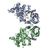
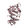
 Assembly
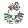


 Mass: 35.453 Da / Num. of mol.: 4 / Source method: obtained synthetically / Formula: Cl
 Mass: 96.063 Da / Num. of mol.: 3 / Source method: obtained synthetically / Formula: SO4
 Mass: 18.015 Da / Num. of mol.: 26 / Source method: isolated from a natural source / Formula: H2O
 Sample preparation
 Processing