 Movie
Movie Controller


+ Open data

- Basic information
| Entry | Database: PDB / ID: 9cip | |||||||||
|---|---|---|---|---|---|---|---|---|---|---|
| Title | MicroED structure of the C11 cysteine protease clostripain | |||||||||
 Components Components | Clostripain | |||||||||
 Keywords Keywords | HYDROLASE / protease / thiol protease | |||||||||
| Function / homology | clostripain / Peptidase C11, Clostripain Clostridium species / Peptidase C11, clostripain / Clostripain family / cysteine-type endopeptidase activity / proteolysis / Clostripain Function and homology information Function and homology information | |||||||||
| Biological species |  Hathewaya histolytica (bacteria) Hathewaya histolytica (bacteria) | |||||||||
| Method | ELECTRON CRYSTALLOGRAPHY / electron crystallography / cryo EM / Resolution: 2.5 Å | |||||||||
 Authors Authors | Ruma, Y.N. / Bu, G. / Hattne, J. / Gonen, T. | |||||||||
| Funding support |  United States, 2items United States, 2items
| |||||||||
 Citation Citation | Journal: J Struct Biol X / Year: 2024 Title: MicroED structure of the C11 cysteine protease clostripain. Authors: Yasmeen N Ruma / Guanhong Bu / Johan Hattne / Tamir Gonen / Abstract: Clostripain secreted from is the founding member of the C11 family of Clan CD cysteine peptidases, which is an important group of peptidases secreted by numerous bacteria. Clostripain is an arginine- ...Clostripain secreted from is the founding member of the C11 family of Clan CD cysteine peptidases, which is an important group of peptidases secreted by numerous bacteria. Clostripain is an arginine-specific endopeptidase. Because of its efficacy as a cysteine peptidase, it is widely used in laboratory settings. Despite its importance the structure of clostripain remains unsolved. Here we describe the first structure of an active form of clostripain determined at 2.5 Å resolution using microcrystal electron diffraction (MicroED). The structure was determined from a single nanocrystal after focused ion beam milling. The structure of clostripain shows a typical Clan CD α/β/α sandwich architecture and the Cys231/His176 catalytic dyad in the active site. It has a large electronegative substrate binding pocket showing its ability to accommodate large and diverse substrates. A loop in the heavy chain formed between residues 452 and 457 is potentially important for substrate binding. In conclusion, this result demonstrates the importance of MicroED to determine the unknown structure of macromolecules such as clostripain, which can be further used as a platform to study substrate binding and design of potential inhibitors against this class of peptidases. | |||||||||
| History |
|
- Structure visualization
| Structure viewer | Molecule: MolmilJmol/JSmol |
|---|
- Downloads & links
-Download
| PDBx/mmCIF format | 9cip.cif.gz | 235.3 KB | Display | PDBx/mmCIF format |
|---|---|---|---|---|
| PDB format | pdb9cip.ent.gz | 148.3 KB | Display | PDB format |
| PDBx/mmJSON format | 9cip.json.gz | Tree view | PDBx/mmJSON format | |
| Others |  Other downloads Other downloads |
-Validation report
| Arichive directory | https://data.pdbj.org/pub/pdb/validation_reports/ci/9cipftp://data.pdbj.org/pub/pdb/validation_reports/ci/9cip | HTTPS FTP |
|---|
-Related structure data
| Related structure data | 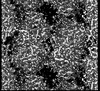 45623MC M: map data used to model this data C: citing same article ( |
|---|---|
| Similar structure data |
-Links
 PDBj
PDBj- Assembly
| Deposited unit | 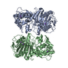
| |||||||||||||||||||||||||||||||||||||||||||||||||||||||||||
|---|---|---|---|---|---|---|---|---|---|---|---|---|---|---|---|---|---|---|---|---|---|---|---|---|---|---|---|---|---|---|---|---|---|---|---|---|---|---|---|---|---|---|---|---|---|---|---|---|---|---|---|---|---|---|---|---|---|---|---|---|
| 1 | 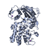
| |||||||||||||||||||||||||||||||||||||||||||||||||||||||||||
| 2 | 
| |||||||||||||||||||||||||||||||||||||||||||||||||||||||||||
| Unit cell |
| |||||||||||||||||||||||||||||||||||||||||||||||||||||||||||
| Noncrystallographic symmetry (NCS) | NCS domain:
NCS domain segments: Ens-ID: ens_1
NCS oper: (Code: givenMatrix: (-0.999998741723, 0.00113373839277, -0.00110959028168), (0.00113802435447, 0.999991865229, -0.00386967381433), (0.00110519405765, -0.00387093168597, -0.999991897184) ...NCS oper: (Code: given Matrix: (-0.999998741723, 0.00113373839277, -0.00110959028168), Vector: |
-Components
| #1: Protein | Mass: 59805.293 Da / Num. of mol.: 2 / Source method: isolated from a natural source / Source: (natural) Hathewaya histolytica (bacteria) / References: UniProt: P09870#2: Chemical | ChemComp-NA /   Mass: 22.990 Da / Num. of mol.: 7 / Source method: obtained synthetically / Formula: Na Mass: 22.990 Da / Num. of mol.: 7 / Source method: obtained synthetically / Formula: Na#3: Water | ChemComp-HOH / |  Mass: 18.015 Da / Num. of mol.: 6 / Source method: isolated from a natural source / Formula: H2O Mass: 18.015 Da / Num. of mol.: 6 / Source method: isolated from a natural source / Formula: H2OHas ligand of interest | N | |
|---|
-Experimental details
-Experiment
| Experiment | Method: ELECTRON CRYSTALLOGRAPHY |
|---|---|
| EM experiment | Aggregation state: 3D ARRAY / 3D reconstruction method: electron crystallography |
- Sample preparation
| Component | Name: Clostripain / Type: COMPLEX / Entity ID: #1 / Source: NATURAL | ||||||||||||||||||||
|---|---|---|---|---|---|---|---|---|---|---|---|---|---|---|---|---|---|---|---|---|---|
| Molecular weight | Value: 0.059 MDa / Experimental value: NO | ||||||||||||||||||||
| Source (natural) | Organism: Hathewaya histolytica (bacteria) | ||||||||||||||||||||
| EM crystal formation | Temperature: 293 K / Time: 1 DAY | ||||||||||||||||||||
| Buffer solution | pH: 5.6 | ||||||||||||||||||||
| Buffer component |
| ||||||||||||||||||||
| Specimen | Embedding applied: NO / Shadowing applied: NO / Staining applied: NO / Vitrification applied: YES | ||||||||||||||||||||
| Specimen support | Grid material: COPPER / Grid mesh size: 200 divisions/in. / Grid type: Quantifoil R2/2 | ||||||||||||||||||||
| Vitrification | Instrument: LEICA EM GP / Cryogen name: ETHANE / Humidity: 95 % / Chamber temperature: 293 K |
-Data collection
| Experimental equipment |  Model: Titan Krios / Image courtesy: FEI Company |
|---|---|
| Microscopy | Model: FEI TITAN KRIOS |
| Electron gun | Electron source:  FIELD EMISSION GUN / Accelerating voltage: 300 kV / Illumination mode: FLOOD BEAM FIELD EMISSION GUN / Accelerating voltage: 300 kV / Illumination mode: FLOOD BEAM |
| Electron lens | Mode: DIFFRACTION / Nominal defocus max: 0 nm / Nominal defocus min: 0 nm |
| Specimen holder | Cryogen: NITROGEN / Temperature (max): 93 K / Temperature (min): 93 K |
| Image recording | Average exposure time: 5 sec. / Electron dose: 0.04 e/Å2 / Film or detector model: FEI FALCON IV (4k x 4k) / Num. of diffraction images: 252 / Num. of grids imaged: 1 / Num. of real images: 252 |
| EM imaging optics | Energyfilter name: TFS Selectris / Energyfilter slit width: 20 eV |
| Image scans | Sampling size: 14 µm / Width: 4096 / Height: 4096 |
| EM diffraction | Camera length: 2500 mm / Tilt angle list: -20,70 |
| EM diffraction shell | Resolution: 2.5→2.56 Å / Fourier space coverage: 86.5 % / Multiplicity: 4.1 / Num. of structure factors: 2318 / Phase residual: 54.5 ° |
| EM diffraction stats | Fourier space coverage: 86.8 % / High resolution: 2.5 Å / Num. of intensities measured: 131476 / Num. of structure factors: 32113 / Phase error: 34.49 ° / Phase error rejection criteria: 0 / Rmerge: 0.279 |
| Reflection | Biso Wilson estimate: 29.39 Å2 |
- Processing
| EM software |
| ||||||||||||||||||||||||||||||||||||||||||||||||||||||||||||||||||||||||||||||||||||
|---|---|---|---|---|---|---|---|---|---|---|---|---|---|---|---|---|---|---|---|---|---|---|---|---|---|---|---|---|---|---|---|---|---|---|---|---|---|---|---|---|---|---|---|---|---|---|---|---|---|---|---|---|---|---|---|---|---|---|---|---|---|---|---|---|---|---|---|---|---|---|---|---|---|---|---|---|---|---|---|---|---|---|---|---|---|
| EM 3D crystal entity | ∠α: 90 ° / ∠β: 90 ° / ∠γ: 90 ° / A: 65.79 Å / B: 106.07 Å / C: 149.28 Å / Space group name: P22121 / Space group num: 18 | ||||||||||||||||||||||||||||||||||||||||||||||||||||||||||||||||||||||||||||||||||||
| CTF correction | Type: NONE | ||||||||||||||||||||||||||||||||||||||||||||||||||||||||||||||||||||||||||||||||||||
| 3D reconstruction | Resolution: 2.5 Å / Resolution method: DIFFRACTION PATTERN/LAYERLINES / Symmetry type: 3D CRYSTAL | ||||||||||||||||||||||||||||||||||||||||||||||||||||||||||||||||||||||||||||||||||||
| Atomic model building | B value: 24.7 / Protocol: FLEXIBLE FIT / Space: RECIPROCAL | ||||||||||||||||||||||||||||||||||||||||||||||||||||||||||||||||||||||||||||||||||||
| Atomic model building | Accession code: A0A4U9RR22 / Source name: AlphaFold / Type: in silico model | ||||||||||||||||||||||||||||||||||||||||||||||||||||||||||||||||||||||||||||||||||||
| Refinement | Resolution: 2.5→49.4 Å / SU ML: 0.3767 / Cross valid method: FREE R-VALUE / Phase error: 34.4948 Stereochemistry target values: GeoStd + Monomer Library + CDL v1.2
| ||||||||||||||||||||||||||||||||||||||||||||||||||||||||||||||||||||||||||||||||||||
| Solvent computation | Shrinkage radii: 0.9 Å / VDW probe radii: 1.1 Å / Solvent model: FLAT BULK SOLVENT MODEL | ||||||||||||||||||||||||||||||||||||||||||||||||||||||||||||||||||||||||||||||||||||
| Displacement parameters | Biso mean: 24.69 Å2 | ||||||||||||||||||||||||||||||||||||||||||||||||||||||||||||||||||||||||||||||||||||
| Refine LS restraints |
| ||||||||||||||||||||||||||||||||||||||||||||||||||||||||||||||||||||||||||||||||||||
| Refine LS restraints NCS | Type: Torsion NCS / Rms dev position: 0.381708976931 Å | ||||||||||||||||||||||||||||||||||||||||||||||||||||||||||||||||||||||||||||||||||||
| LS refinement shell |
|
