 ムービー
ムービー コントローラー


+ データを開く

- 基本情報
基本情報
| 登録情報 | データベース: PDB / ID: 9cgi | ||||||
|---|---|---|---|---|---|---|---|
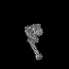
| タイトル | Cryo-EM structure of the Nipah Virus polymerase (L) protein in complex with the tetrameric phosphoprotein (P) | ||||||
 要素 要素 |
| ||||||
 キーワード キーワード | TRANSFERASE / VIRAL PROTEIN / Nipah virus / L protein / phosphoprotein / RNA-dependent RNA polymerase / PRNTase / GDP polyribonucleotidyl transferase / RNA capping / viral replication | ||||||
| 機能・相同性 |  機能・相同性情報 機能・相同性情報negative stranded viral RNA transcription / NNS virus cap methyltransferase / GDP polyribonucleotidyltransferase / negative stranded viral RNA replication / 加水分解酵素; 酸無水物に作用; リン含有酸無水物に作用 / virion component / molecular adaptor activity / host cell cytoplasm / mRNA 5'-cap (guanine-N7-)-methyltransferase activity / symbiont-mediated suppression of host innate immune response ...negative stranded viral RNA transcription / NNS virus cap methyltransferase / GDP polyribonucleotidyltransferase / negative stranded viral RNA replication / 加水分解酵素; 酸無水物に作用; リン含有酸無水物に作用 / virion component / molecular adaptor activity / host cell cytoplasm / mRNA 5'-cap (guanine-N7-)-methyltransferase activity / symbiont-mediated suppression of host innate immune response / RNA-directed RNA polymerase / RNA-directed RNA polymerase activity / GTPase activity / ATP binding 類似検索 - 分子機能 | ||||||
| 生物種 |  Henipavirus nipahense (ウイルス) Henipavirus nipahense (ウイルス) | ||||||
| 手法 | 電子顕微鏡法 / 単粒子再構成法 / クライオ電子顕微鏡法 / 解像度: 2.92 Å | ||||||
 データ登録者 データ登録者 | Liu, B. / Yang, G. / Wang, D. | ||||||
| 資金援助 | 1件
| ||||||
 引用 引用 | ジャーナル: Nat Commun / 年: 2024 タイトル: Structure of the Nipah virus polymerase phosphoprotein complex. 著者: Ge Yang / Dong Wang / Bin Liu /  要旨: The Nipah virus (NiV), a member of the Paramyxoviridae family, is notorious for its high fatality rate in humans. The RNA polymerase machinery of NiV, comprising the large protein L and the ...The Nipah virus (NiV), a member of the Paramyxoviridae family, is notorious for its high fatality rate in humans. The RNA polymerase machinery of NiV, comprising the large protein L and the phosphoprotein P, is essential for viral replication. This study presents the 2.9-Å cryo-electron microscopy structure of the NiV L-P complex, shedding light on its assembly and functionality. The structure not only demonstrates the molecular details of the conserved N-terminal domain, RNA-dependent RNA polymerase (RdRp), and GDP polyribonucleotidyltransferase of the L protein, but also the intact central oligomerization domain and the C-terminal X domain of the P protein. The P protein interacts extensively with the L protein, forming an antiparallel β-sheet among the P protomers and with the fingers subdomain of RdRp. The flexible linker domain of one P promoter extends its contact with the fingers subdomain to reach near the nascent RNA exit, highlighting the distinct characteristic of the NiV L-P interface. This distinctive tetrameric organization of the P protein and its interaction with the L protein provide crucial molecular insights into the replication and transcription mechanisms of NiV polymerase, ultimately contributing to the development of effective treatments and preventive measures against this Paramyxoviridae family deadly pathogen. | ||||||
| 履歴 |
|
- 構造の表示
構造の表示
| 構造ビューア | 分子: MolmilJmol/JSmol |
|---|
- ダウンロードとリンク
-ダウンロード
| PDBx/mmCIF形式 | 9cgi.cif.gz | 422.3 KB | 表示 | PDBx/mmCIF形式 |
|---|---|---|---|---|
| PDB形式 | pdb9cgi.ent.gz | 311.5 KB | 表示 | PDB形式 |
| PDBx/mmJSON形式 | 9cgi.json.gz | ツリー表示 | PDBx/mmJSON形式 | |
| その他 |  その他のダウンロード その他のダウンロード |
-検証レポート
| 文書・要旨 | 9cgi_validation.pdf.gz | 1.2 MB | 表示 | wwPDB検証レポート |
|---|---|---|---|---|
| 文書・詳細版 | 9cgi_full_validation.pdf.gz | 1.2 MB | 表示 | |
| XML形式データ | 9cgi_validation.xml.gz | 62.4 KB | 表示 | |
| CIF形式データ | 9cgi_validation.cif.gz | 93.9 KB | 表示 | |
| アーカイブディレクトリ | https://data.pdbj.org/pub/pdb/validation_reports/cg/9cgiftp://data.pdbj.org/pub/pdb/validation_reports/cg/9cgi | HTTPS FTP |
-関連構造データ
-リンク
 PDBj
PDBj


- 集合体
集合体
| 登録構造単位 | 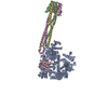
|
|---|---|
| 1 |
|
-要素
| #1: タンパク質 | 分子量: 257565.156 Da / 分子数: 1 / 由来タイプ: 組換発現 / 由来: (組換発現) Henipavirus nipahense (ウイルス) / 細胞株 (発現宿主): Sf9発現宿主:   Spodoptera frugiperda (ツマジロクサヨトウ) Spodoptera frugiperda (ツマジロクサヨトウ)参照: UniProt: Q997F0, RNA-directed RNA polymerase, 加水分解酵素; 酸無水物に作用; リン含有酸無水物に作用, GDP polyribonucleotidyltransferase, NNS virus cap methyltransferase | ||
|---|---|---|---|
| #2: タンパク質 | 分子量: 78390.320 Da / 分子数: 4 / 由来タイプ: 組換発現 / 由来: (組換発現) Henipavirus nipahense (ウイルス) / 遺伝子: P/V/C / 細胞株 (発現宿主): Sf9発現宿主: Spodoptera frugiperda (ツマジロクサヨトウ)参照: UniProt: Q9IK91 Has protein modification | Y | |
-実験情報
-実験
| 実験 | 手法: 電子顕微鏡法 |
|---|---|
| EM実験 | 試料の集合状態: PARTICLE / 3次元再構成法: 単粒子再構成法 |
- 試料調製
試料調製
| 構成要素 | 名称: The Nipah virus L-P complex / タイプ: COMPLEX / Entity ID: all / 由来: RECOMBINANT |
|---|---|
| 分子量 | 値: 0.65 MDa / 実験値: YES |
| 由来(天然) | 生物種: Henipavirus nipahense (ウイルス) |
| 由来(組換発現) | 生物種: Spodoptera frugiperda (ツマジロクサヨトウ) |
| 緩衝液 | pH: 8 詳細: 50 mM Tris-HCl pH 8.0, 250 mM NaCl, 5% glycerol, 1 mM TCEP, and 4 mM MgCl2 |
| 試料 | 包埋: NO / シャドウイング: NO / 染色: NO / 凍結: YES |
| 試料支持 | グリッドの材料: COPPER / グリッドのサイズ: 300 divisions/in. / グリッドのタイプ: Quantifoil R1.2/1.3 |
| 急速凍結 | 装置: FEI VITROBOT MARK IV / 凍結剤: ETHANE / 湿度: 100 % / 凍結前の試料温度: 277 K |
- 電子顕微鏡撮影
| 実験機器 |  モデル: Titan Krios / 画像提供: FEI Company |
|---|---|
| 顕微鏡 | モデル: FEI TITAN KRIOS |
| 電子銃 | 電子線源:  FIELD EMISSION GUN / 加速電圧: 300 kV / 照射モード: FLOOD BEAM FIELD EMISSION GUN / 加速電圧: 300 kV / 照射モード: FLOOD BEAM |
| 電子レンズ | モード: BRIGHT FIELD / 倍率(公称値): 130000 X / 最大 デフォーカス(公称値): 2000 nm / 最小 デフォーカス(公称値): 1000 nm / Cs: 2.7 mm / C2レンズ絞り径: 50 µm / アライメント法: COMA FREE |
| 試料ホルダ | 凍結剤: NITROGEN 試料ホルダーモデル: FEI TITAN KRIOS AUTOGRID HOLDER 最高温度: 77 K / 最低温度: 63 K |
| 撮影 | 平均露光時間: 1.7 sec. / 電子線照射量: 54.8 e/Å2 フィルム・検出器のモデル: GATAN K3 BIOQUANTUM (6k x 4k) 撮影したグリッド数: 1 / 実像数: 6114 |
| 画像スキャン | 横: 5760 / 縦: 4092 |
- 解析
解析
| EMソフトウェア | 名称: PHENIX / バージョン: 1.21_5207: / カテゴリ: モデル精密化 | ||||||||||||||||||||||||
|---|---|---|---|---|---|---|---|---|---|---|---|---|---|---|---|---|---|---|---|---|---|---|---|---|---|
| CTF補正 | タイプ: PHASE FLIPPING AND AMPLITUDE CORRECTION | ||||||||||||||||||||||||
| 粒子像の選択 | 選択した粒子像数: 3052781 | ||||||||||||||||||||||||
| 対称性 | 点対称性: C1 (非対称) | ||||||||||||||||||||||||
| 3次元再構成 | 解像度: 2.92 Å / 解像度の算出法: FSC 0.143 CUT-OFF / 粒子像の数: 278295 / アルゴリズム: SIMULTANEOUS ITERATIVE (SIRT) / クラス平均像の数: 1 / 対称性のタイプ: POINT | ||||||||||||||||||||||||
| 原子モデル構築 | B value: 73.5 / プロトコル: OTHER / 空間: REAL | ||||||||||||||||||||||||
| 拘束条件 |
|
