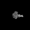
 Movie
Movie Controller

[English] 日本語
 Yorodumi
- PDB-9cfw: Cryo-EM structure of myosin-1c bound to F-actin in the ADP-B state -
+ Open data

- Basic information
| Entry | Database: PDB / ID: 9cfw | |||||||||
|---|---|---|---|---|---|---|---|---|---|---|
| Title | Cryo-EM structure of myosin-1c bound to F-actin in the ADP-B state | |||||||||
 Components Components |
| |||||||||
 Keywords Keywords | MOTOR PROTEIN / F-actin / myosin / myosin-1c / cellular motility / cryo-EM / actomyosin | |||||||||
| Function / homology |  Function and homology information Function and homology informationstereocilium membrane / B-WICH complex positively regulates rRNA expression / positive regulation of cellular response to insulin stimulus / CaMK IV-mediated phosphorylation of CREB / Cam-PDE 1 activation / CREB1 phosphorylation through the activation of CaMKII/CaMKK/CaMKIV cascasde / Glycogen breakdown (glycogenolysis) / Activation of RAC1 downstream of NMDARs / Sodium/Calcium exchangers / Activation of Ca-permeable Kainate Receptor ...stereocilium membrane / B-WICH complex positively regulates rRNA expression / positive regulation of cellular response to insulin stimulus / CaMK IV-mediated phosphorylation of CREB / Cam-PDE 1 activation / CREB1 phosphorylation through the activation of CaMKII/CaMKK/CaMKIV cascasde / Glycogen breakdown (glycogenolysis) / Activation of RAC1 downstream of NMDARs / Sodium/Calcium exchangers / Activation of Ca-permeable Kainate Receptor / CLEC7A (Dectin-1) induces NFAT activation / RHO GTPases activate PAKs / Calmodulin induced events / Synthesis of IP3 and IP4 in the cytosol / Inactivation, recovery and regulation of the phototransduction cascade / Tetrahydrobiopterin (BH4) synthesis, recycling, salvage and regulation / eNOS activation / Reduction of cytosolic Ca++ levels / Calcineurin activates NFAT / Ion transport by P-type ATPases / RAF activation / VEGFR2 mediated vascular permeability / Protein methylation / vesicle transport along actin filament / RAS processing / Ca2+ pathway / FCERI mediated Ca+2 mobilization / Extra-nuclear estrogen signaling / RHO GTPases activate IQGAPs / Unblocking of NMDA receptors, glutamate binding and activation / PKA activation / RAF/MAP kinase cascade / Regulation of actin dynamics for phagocytic cup formation / Smooth Muscle Contraction / Platelet degranulation / stereocilium bundle / High laminar flow shear stress activates signaling by PIEZO1 and PECAM1:CDH5:KDR in endothelial cells / actin filament-based movement / B-WICH complex / Stimuli-sensing channels / : / Ion homeostasis / type 3 metabotropic glutamate receptor binding / stereocilium / myosin complex / protein targeting to membrane / positive regulation of transcription by RNA polymerase III / regulation of bicellular tight junction assembly / cytoskeletal motor activator activity / vascular endothelial growth factor signaling pathway / microfilament motor activity / response to corticosterone / myosin heavy chain binding / negative regulation of ryanodine-sensitive calcium-release channel activity / organelle localization by membrane tethering / : / autophagosome membrane docking / regulation of synaptic vesicle exocytosis / regulation of ryanodine-sensitive calcium-release channel activity / regulation of cardiac muscle cell action potential / tropomyosin binding / presynaptic endocytosis / actin filament bundle / troponin I binding / positive regulation of transcription by RNA polymerase I / filamentous actin / mesenchyme migration / microvillus / positive regulation of actin filament polymerization / calcineurin-mediated signaling / nitric-oxide synthase binding / regulation of cell communication by electrical coupling involved in cardiac conduction / skeletal muscle myofibril / brush border / actin filament bundle assembly / striated muscle thin filament / adenylate cyclase binding / protein phosphatase activator activity / skeletal muscle thin filament assembly / lateral plasma membrane / actin monomer binding / positive regulation of protein targeting to membrane / regulation of synaptic vesicle endocytosis / detection of calcium ion / postsynaptic cytosol / regulation of cardiac muscle contraction / cell surface receptor signaling pathway via JAK-STAT / catalytic complex / phosphatidylinositol 3-kinase binding / calcium channel inhibitor activity / presynaptic cytosol / skeletal muscle fiber development / cellular response to interferon-beta / phagocytic vesicle / regulation of release of sequestered calcium ion into cytosol by sarcoplasmic reticulum / stress fiber / titin binding / actin filament polymerization / regulation of cardiac muscle contraction by regulation of the release of sequestered calcium ion / voltage-gated potassium channel complex Similarity search - Function | |||||||||
| Biological species |  | |||||||||
| Method | ELECTRON MICROSCOPY / helical reconstruction / cryo EM / Resolution: 3 Å | |||||||||
 Authors Authors | Chavali, S.S. / Sindelar, C.V. / Ostap, M.E. | |||||||||
| Funding support |  United States, 2items United States, 2items
| |||||||||
 Citation Citation | Journal: Proc Natl Acad Sci U S A / Year: 2025 Title: High-resolution structures of Myosin-IC reveal a unique actin-binding orientation, ADP release pathway, and power stroke trajectory. Authors: Sai Shashank Chavali / Peter J Carman / Henry Shuman / E Michael Ostap / Charles V Sindelar / Abstract: Myosin-IC (myo1c) is a class-I myosin that supports transport and remodeling of the plasma membrane and membrane-bound vesicles. Like other members of the myosin family, its biochemical kinetics are ...Myosin-IC (myo1c) is a class-I myosin that supports transport and remodeling of the plasma membrane and membrane-bound vesicles. Like other members of the myosin family, its biochemical kinetics are altered in response to changes in mechanical loads that resist the power stroke. However, myo1c is unique in that the primary force-sensitive kinetic transition is the isomerization that follows ATP binding, not ADP release as in other slow myosins. Myo1c also powers actin gliding along curved paths, propelling actin filaments in leftward circles. To understand the origins of this unique force-sensing and motile behavior, we solved actin-bound myo1c cryo-EM structures in the presence and absence of ADP. Our structures reveal that in contrast with other myosins, the myo1c lever arm swing is skewed, partly due to a different actin interface that reorients the motor domain on actin. The structures also reveal unique nucleotide-dependent behavior of both the nucleotide pocket as well as an element called the N-terminal extension (NTE). We incorporate these observations into a model that explains why force primarily regulates ATP binding in myo1c, rather than ADP release as in other myosins. Integrating our cryo-EM data with available crystallography structures allows the modeling of full-length myo1c during force generation, supplying insights into its role in membrane remodeling. These results highlight how relatively minor sequence differences in members of the myosin superfamily can significantly alter power stroke geometry and force-sensing properties, with important implications for biological function. | |||||||||
| History |
|
- Structure visualization
| Structure viewer | Molecule: MolmilJmol/JSmol |
|---|
- Downloads & links
-Download
| PDBx/mmCIF format | 9cfw.cif.gz | 362.5 KB | Display | PDBx/mmCIF format |
|---|---|---|---|---|
| PDB format | pdb9cfw.ent.gz | 292.7 KB | Display | PDB format |
| PDBx/mmJSON format | 9cfw.json.gz | Tree view | PDBx/mmJSON format | |
| Others |  Other downloads Other downloads |
-Validation report
| Arichive directory | https://data.pdbj.org/pub/pdb/validation_reports/cf/9cfwftp://data.pdbj.org/pub/pdb/validation_reports/cf/9cfw | HTTPS FTP |
|---|
-Related structure data
| Related structure data |  45565MC  9cfuC  9cfvC  9cfxC M: map data used to model this data C: citing same article ( |
|---|---|
| Similar structure data |


-Links
 PDBj
PDBj


- Assembly
| Deposited unit | 
|
|---|---|
| 1 |
|
-Components
| #1: Protein | Mass: 41862.613 Da / Num. of mol.: 3 / Source method: isolated from a natural source / Source: (natural) References: UniProt: P68135, Hydrolases; Acting on acid anhydrides; Acting on acid anhydrides to facilitate cellular and subcellular movement #2: Protein | | Mass: 92064.203 Da / Num. of mol.: 1 Source method: isolated from a genetically manipulated source Source: (gene. exp.)  unidentified baculovirus / References: UniProt: Q9WTI7 unidentified baculovirus / References: UniProt: Q9WTI7#3: Protein | | Mass: 16723.365 Da / Num. of mol.: 1 Source method: isolated from a genetically manipulated source Source: (gene. exp.) unidentified baculovirus / References: UniProt: P0DP26#4: Chemical | ChemComp-ADP /   Mass: 427.201 Da / Num. of mol.: 4 / Source method: obtained synthetically / Formula: C10H15N5O10P2 / Comment: ADP, energy-carrying molecule*YM Mass: 427.201 Da / Num. of mol.: 4 / Source method: obtained synthetically / Formula: C10H15N5O10P2 / Comment: ADP, energy-carrying molecule*YM#5: Chemical |   Mass: 24.305 Da / Num. of mol.: 3 / Source method: obtained synthetically / Formula: Mg Mass: 24.305 Da / Num. of mol.: 3 / Source method: obtained synthetically / Formula: MgHas ligand of interest | N | Has protein modification | N | |
|---|
-Experimental details
-Experiment
| Experiment | Method: ELECTRON MICROSCOPY |
|---|---|
| EM experiment | Aggregation state: FILAMENT / 3D reconstruction method: helical reconstruction |
- Sample preparation
| Component | Name: Complex of myosin-1c with F-actin / Type: COMPLEX / Entity ID: #1-#3 / Source: RECOMBINANT |
|---|---|
| Source (natural) | Organism: |
| Source (recombinant) | Organism: unidentified baculovirus |
| Buffer solution | pH: 7 |
| Specimen | Embedding applied: NO / Shadowing applied: NO / Staining applied: NO / Vitrification applied: YES |
| Vitrification | Cryogen name: ETHANE |
- Electron microscopy imaging
| Experimental equipment |  Model: Titan Krios / Image courtesy: FEI Company |
|---|---|
| Microscopy | Model: TFS KRIOS |
| Electron gun | Electron source:  FIELD EMISSION GUN / Accelerating voltage: 300 kV / Illumination mode: OTHER FIELD EMISSION GUN / Accelerating voltage: 300 kV / Illumination mode: OTHER |
| Electron lens | Mode: BRIGHT FIELD / Nominal defocus max: 2500 nm / Nominal defocus min: 1200 nm |
| Image recording | Electron dose: 52 e/Å2 / Film or detector model: GATAN K3 (6k x 4k) |
- Processing
| CTF correction | Type: PHASE FLIPPING AND AMPLITUDE CORRECTION |
|---|---|
| Helical symmerty | Angular rotation/subunit: 167.88 ° / Axial rise/subunit: 27.38 Å / Axial symmetry: C1 |
| 3D reconstruction | Resolution: 3 Å / Resolution method: FSC 0.143 CUT-OFF / Num. of particles: 126845 / Symmetry type: HELICAL |
