 Movie
Movie Controller

[English] 日本語
 Yorodumi
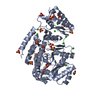
- PDB-9c9o: M. tuberculosis PKS13 acyltransferase (AT) domain in complex with... -
+ Open data

- Basic information
| Entry | Database: PDB / ID: 9c9o | ||||||||||||
|---|---|---|---|---|---|---|---|---|---|---|---|---|---|
| Title | M. tuberculosis PKS13 acyltransferase (AT) domain in complex with SuFEx inhibitor CMX410 - reaction product | ||||||||||||
 Components Components | (Polyketide synthase ...) x 2 | ||||||||||||
 Keywords Keywords | TRANSFERASE / polyketide synthase / acyltransferase domain / inhibitor / mycobacteria / Structural Genomics / TB Structural Genomics Consortium / TBSGC | ||||||||||||
| Function / homology |  Function and homology information Function and homology informationDIM/DIP cell wall layer assembly / fatty acid synthase activity / phosphopantetheine binding / 3-oxoacyl-[acyl-carrier-protein] synthase activity / Transferases; Acyltransferases; Transferring groups other than aminoacyl groups / fatty acid biosynthetic process / cytoplasm Similarity search - Function | ||||||||||||
| Biological species |   Mycobacterium tuberculosis (bacteria) Mycobacterium tuberculosis (bacteria) | ||||||||||||
| Method |  X-RAY DIFFRACTION / SYNCHROTRON / MOLECULAR REPLACEMENT / Resolution: 2.02 Å X-RAY DIFFRACTION / SYNCHROTRON / MOLECULAR REPLACEMENT / Resolution: 2.02 Å | ||||||||||||
 Authors Authors | Krieger, I.V. / Tang, S. / Sacchetini, J.C. / TB Structural Genomics Consortium (TBSGC) | ||||||||||||
| Funding support |  United States, 3items United States, 3items
| ||||||||||||
 Citation Citation | Journal: Nature / Year: 2025 Title: SuFEx-based antitubercular compound irreversibly inhibits Pks13. Authors: Krieger, I.V. / Sukheja, P. / Yang, B. / Tang, S. / Selle, D. / Woods, A. / Engelhart, C. / Kumar, P. / Harbut, M.B. / Liu, D. / Tsuda, B. / Qin, B. / Bare, G.A.L. / Li, G. / Chi, V. / ...Authors: Krieger, I.V. / Sukheja, P. / Yang, B. / Tang, S. / Selle, D. / Woods, A. / Engelhart, C. / Kumar, P. / Harbut, M.B. / Liu, D. / Tsuda, B. / Qin, B. / Bare, G.A.L. / Li, G. / Chi, V. / Gambacurta, J. / Hvizdos, J. / Reagan, M. / Jones, I.L. / Massoudi, L.M. / Woolhiser, L.K. / Cascioferro, A. / Kundrick, E. / Singh, P. / Reiley, W. / Ioerger, T.R. / Kandula, D.R. / McCabe, J.W. / Guo, T. / Alland, D. / Boshoff, H.I. / Schnappinger, D. / Robertson, G.T. / Mdluli, K. / Lee, K.J. / Dong, J. / Li, S. / Schultz, P.G. / Joseph, S.B. / Love, M.S. / Sharpless, K.B. / Petrassi, H.M. / Chatterjee, A.K. / Sacchettini, J.C. / McNamara, C.W. | ||||||||||||
| History |
|
- Structure visualization
| Structure viewer | Molecule: MolmilJmol/JSmol |
|---|
- Downloads & links
-Download
| PDBx/mmCIF format | 9c9o.cif.gz | 269.2 KB | Display | PDBx/mmCIF format |
|---|---|---|---|---|
| PDB format | pdb9c9o.ent.gz | Display | PDB format | |
| PDBx/mmJSON format | 9c9o.json.gz | Tree view | PDBx/mmJSON format | |
| Others |  Other downloads Other downloads |
-Validation report
| Arichive directory | https://data.pdbj.org/pub/pdb/validation_reports/c9/9c9oftp://data.pdbj.org/pub/pdb/validation_reports/c9/9c9o | HTTPS FTP |
|---|
-Related structure data


-Links
 PDBj
PDBj


- Assembly
| Deposited unit | 
| ||||||||||||
|---|---|---|---|---|---|---|---|---|---|---|---|---|---|
| 1 | 
| ||||||||||||
| 2 | 
| ||||||||||||
| 3 | 
| ||||||||||||
| Unit cell |
| ||||||||||||
| Components on special symmetry positions |
|
-Components
-Polyketide synthase ... , 2 types, 3 molecules ABC
| #1: Protein | Mass: 55527.441 Da / Num. of mol.: 1 Source method: isolated from a genetically manipulated source Source: (gene. exp.) Mycobacterium tuberculosis (strain ATCC 25618 / H37Rv) (bacteria)Strain: ATCC 25618 / H37Rv / Gene: pks13, Rv3800c / Production host: References: UniProt: I6X8D2, Transferases; Acyltransferases; Transferring groups other than aminoacyl groups |
|---|---|
| #2: Protein | Mass: 55546.441 Da / Num. of mol.: 2 Source method: isolated from a genetically manipulated source Source: (gene. exp.) Mycobacterium tuberculosis (strain ATCC 25618 / H37Rv) (bacteria)Strain: ATCC 25618 / H37Rv / Gene: pks13, Rv3800c / Production host: References: UniProt: I6X8D2, Transferases; Acyltransferases; Transferring groups other than aminoacyl groups |
-Sugars , 1 types, 7 molecules
| #3: Polysaccharide | beta-D-fructofuranose-(2-1)-alpha-D-glucopyranose   Source method: isolated from a genetically manipulated source Details: oligosaccharide with reducing-end-to-reducing-end glycosidic bond References: sucrose |
|---|
-Non-polymers , 4 types, 451 molecules 


| #4: Chemical | ChemComp-SO4 /  Mass: 96.063 Da / Num. of mol.: 4 / Source method: obtained synthetically / Formula: SO4 Mass: 96.063 Da / Num. of mol.: 4 / Source method: obtained synthetically / Formula: SO4#5: Chemical |  Mass: 238.278 Da / Num. of mol.: 2 / Source method: obtained synthetically / Formula: C10H22O6 / Comment: precipitant*YM Mass: 238.278 Da / Num. of mol.: 2 / Source method: obtained synthetically / Formula: C10H22O6 / Comment: precipitant*YM#6: Chemical | ChemComp-A1AVL / | Mass: 476.432 Da / Num. of mol.: 1 / Source method: obtained synthetically / Formula: C16H14F2N4O7S2 / Feature type: SUBJECT OF INVESTIGATION #7: Water | ChemComp-HOH / | Mass: 18.015 Da / Num. of mol.: 444 / Source method: isolated from a natural source / Formula: H2O |
|---|
-Details
| Compound details | The authors state that the chain C short peptide is part of the ordered N-terminal part of either ...The authors state that the chain C short peptide is part of the ordered N-terminal part of either chain A or chain B, but because of how much is missing, they cannot say whether is it from chain A or chain B. |
|---|---|
| Has ligand of interest | Y |
| Has protein modification | Y |
-Experimental details
-Experiment
| Experiment | Method: X-RAY DIFFRACTION / Number of used crystals: 1 |
|---|
- Sample preparation
| Crystal | Density Matthews: 3.59 Å3/Da / Density % sol: 65.78 % |
|---|---|
| Crystal grow | Temperature: 290 K / Method: vapor diffusion, hanging drop / Details: 0.1 M HEPES pH 7.5, 1.7 M (NH4)2SO4, 4-6% PEG 400 |
-Data collection
| Diffraction | Mean temperature: 120 K / Serial crystal experiment: N |
|---|---|
| Diffraction source | Source: SYNCHROTRON / Site: NSLS-II / Beamline: 17-ID-1 / Wavelength: 0.920105 Å |
| Detector | Type: DECTRIS EIGER X 9M / Detector: PIXEL / Date: Sep 26, 2022 |
| Radiation | Protocol: SINGLE WAVELENGTH / Monochromatic (M) / Laue (L): M / Scattering type: x-ray |
| Radiation wavelength | Wavelength: 0.920105 Å / Relative weight: 1 |
| Reflection | Resolution: 2.019→98.62 Å / Num. obs: 79459 / % possible obs: 92.6 % / Redundancy: 7 % / Biso Wilson estimate: 31.34 Å2 / CC1/2: 0.997 / Rmerge(I) obs: 0.126 / Rpim(I) all: 0.051 / Net I/σ(I): 10.5 |
| Reflection shell | Resolution: 2.019→2.163 Å / Redundancy: 6.5 % / Rmerge(I) obs: 1.324 / Num. unique obs: 3973 / CC1/2: 0.579 / Rpim(I) all: 0.561 / % possible all: 67.1 |
- Processing
| Software |
| |||||||||||||||||||||||||||||||||||||||||||||||||||||||||||||||||||||||||||||||||||||||||||||||||||||||||||||||||||||||||||||||||||||||||||||||||||||||||||||||||||||||||||||||||||||||||||||||||||||||||||
|---|---|---|---|---|---|---|---|---|---|---|---|---|---|---|---|---|---|---|---|---|---|---|---|---|---|---|---|---|---|---|---|---|---|---|---|---|---|---|---|---|---|---|---|---|---|---|---|---|---|---|---|---|---|---|---|---|---|---|---|---|---|---|---|---|---|---|---|---|---|---|---|---|---|---|---|---|---|---|---|---|---|---|---|---|---|---|---|---|---|---|---|---|---|---|---|---|---|---|---|---|---|---|---|---|---|---|---|---|---|---|---|---|---|---|---|---|---|---|---|---|---|---|---|---|---|---|---|---|---|---|---|---|---|---|---|---|---|---|---|---|---|---|---|---|---|---|---|---|---|---|---|---|---|---|---|---|---|---|---|---|---|---|---|---|---|---|---|---|---|---|---|---|---|---|---|---|---|---|---|---|---|---|---|---|---|---|---|---|---|---|---|---|---|---|---|---|---|---|---|---|---|---|---|---|
| Refinement | Method to determine structure: MOLECULAR REPLACEMENT / Resolution: 2.02→52.24 Å / SU ML: 0.2437 / Cross valid method: FREE R-VALUE / σ(F): 1.34 / Phase error: 25.5167 Stereochemistry target values: GeoStd + Monomer Library + CDL v1.2
| |||||||||||||||||||||||||||||||||||||||||||||||||||||||||||||||||||||||||||||||||||||||||||||||||||||||||||||||||||||||||||||||||||||||||||||||||||||||||||||||||||||||||||||||||||||||||||||||||||||||||||
| Solvent computation | Shrinkage radii: 0.9 Å / VDW probe radii: 1.11 Å / Solvent model: FLAT BULK SOLVENT MODEL | |||||||||||||||||||||||||||||||||||||||||||||||||||||||||||||||||||||||||||||||||||||||||||||||||||||||||||||||||||||||||||||||||||||||||||||||||||||||||||||||||||||||||||||||||||||||||||||||||||||||||||
| Displacement parameters | Biso mean: 35.25 Å2 | |||||||||||||||||||||||||||||||||||||||||||||||||||||||||||||||||||||||||||||||||||||||||||||||||||||||||||||||||||||||||||||||||||||||||||||||||||||||||||||||||||||||||||||||||||||||||||||||||||||||||||
| Refinement step | Cycle: LAST / Resolution: 2.02→52.24 Å
| |||||||||||||||||||||||||||||||||||||||||||||||||||||||||||||||||||||||||||||||||||||||||||||||||||||||||||||||||||||||||||||||||||||||||||||||||||||||||||||||||||||||||||||||||||||||||||||||||||||||||||
| Refine LS restraints |
| |||||||||||||||||||||||||||||||||||||||||||||||||||||||||||||||||||||||||||||||||||||||||||||||||||||||||||||||||||||||||||||||||||||||||||||||||||||||||||||||||||||||||||||||||||||||||||||||||||||||||||
| LS refinement shell |
|
