 Movie
Movie Controller


+ Open data

- Basic information
| Entry | Database: PDB / ID: 9c8j | ||||||
|---|---|---|---|---|---|---|---|
| Title | X-ray crystal structure of AmpC beta-lactamase with inhibitor | ||||||
 Components Components | AmpC Beta-lactamase | ||||||
 Keywords Keywords | HYDROLASE / inhibitor | ||||||
| Function / homology |  Function and homology information Function and homology informationantibiotic catabolic process / beta-lactamase activity / beta-lactamase / outer membrane-bounded periplasmic space / response to antibiotic Similarity search - Function | ||||||
| Biological species |  | ||||||
| Method |  X-RAY DIFFRACTION / SYNCHROTRON / MOLECULAR REPLACEMENT / Resolution: 1.55 Å X-RAY DIFFRACTION / SYNCHROTRON / MOLECULAR REPLACEMENT / Resolution: 1.55 Å | ||||||
 Authors Authors | Liu, F. / Shoichet, B.K. | ||||||
| Funding support |  United States, 1items United States, 1items
| ||||||
 Citation Citation | Journal: To Be Published Title: Improved correlations with score, hit-rate, and affinity as docking library and testing scale increase Authors: Liu, F. / Shoichet, B.K. | ||||||
| History |
|
- Structure visualization
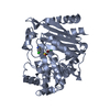
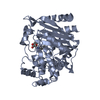
| Structure viewer | Molecule: MolmilJmol/JSmol |
|---|
- Downloads & links
-Download
| PDBx/mmCIF format | 9c8j.cif.gz | 184 KB | Display | PDBx/mmCIF format |
|---|---|---|---|---|
| PDB format | pdb9c8j.ent.gz | Display | PDB format | |
| PDBx/mmJSON format | 9c8j.json.gz | Tree view | PDBx/mmJSON format | |
| Others |  Other downloads Other downloads |
-Validation report
| Summary document | 9c8j_validation.pdf.gz | 1.2 MB | Display | wwPDB validaton report |
|---|---|---|---|---|
| Full document | 9c8j_full_validation.pdf.gz | 1.2 MB | Display | |
| Data in XML | 9c8j_validation.xml.gz | 33.6 KB | Display | |
| Data in CIF | 9c8j_validation.cif.gz | 50.3 KB | Display | |
| Arichive directory | https://data.pdbj.org/pub/pdb/validation_reports/c8/9c8jftp://data.pdbj.org/pub/pdb/validation_reports/c8/9c8j | HTTPS FTP |
-Related structure data
-Links
 PDBj
PDBj
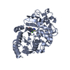
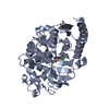
- Assembly
| Deposited unit | 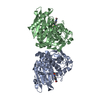
| ||||||||||
|---|---|---|---|---|---|---|---|---|---|---|---|
| 1 | 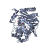
| ||||||||||
| 2 | 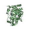
| ||||||||||
| Unit cell |
|
-Components
| #1: Protein | Mass: 41593.359 Da / Num. of mol.: 2 Source method: isolated from a genetically manipulated source Source: (gene. exp.) #2: Chemical | Mass: 366.819 Da / Num. of mol.: 2 / Source method: isolated from a natural source / Formula: C16H15ClN2O4S / Feature type: SUBJECT OF INVESTIGATION #3: Water | ChemComp-HOH / |  Mass: 18.015 Da / Num. of mol.: 608 / Source method: isolated from a natural source / Formula: H2O Mass: 18.015 Da / Num. of mol.: 608 / Source method: isolated from a natural source / Formula: H2OHas ligand of interest | Y | |
|---|
-Experimental details
-Experiment
| Experiment | Method: X-RAY DIFFRACTION / Number of used crystals: 1 |
|---|
- Sample preparation
| Crystal | Density Matthews: 2.48 Å3/Da / Density % sol: 50.5 % |
|---|---|
| Crystal grow | Temperature: 293.15 K / Method: vapor diffusion, hanging drop / Details: 2M KPI; pH 8.56 |
-Data collection
| Diffraction | Mean temperature: 100 K / Serial crystal experiment: N |
|---|---|
| Diffraction source | Source: SYNCHROTRON / Site: ALS / Beamline: 8.3.1 / Wavelength: 1.11583 Å |
| Detector | Type: DECTRIS PILATUS3 S 6M / Detector: PIXEL / Date: Oct 21, 2023 |
| Radiation | Protocol: MAD / Monochromatic (M) / Laue (L): M / Scattering type: x-ray |
| Radiation wavelength | Wavelength: 1.11583 Å / Relative weight: 1 |
| Reflection | Resolution: 1.55→58.45 Å / Num. obs: 112725 / % possible obs: 99.31 % / Redundancy: 1.99 % / Biso Wilson estimate: 23.36 Å2 / CC1/2: 0.999 / CC star: 1 / Rmerge(I) obs: 0.02917 / Net I/σ(I): 14.17 |
| Reflection shell | Resolution: 1.55→1.605 Å / Redundancy: 1.99 % / Rmerge(I) obs: 0.8336 / Mean I/σ(I) obs: 1.05 / Num. unique obs: 10870 / CC1/2: 0.439 / CC star: 0.781 / % possible all: 95.75 |
- Processing
| Software |
| |||||||||||||||||||||||||||||||||||||||||||||||||||||||||||||||||||||||||||||||||||||||||||
|---|---|---|---|---|---|---|---|---|---|---|---|---|---|---|---|---|---|---|---|---|---|---|---|---|---|---|---|---|---|---|---|---|---|---|---|---|---|---|---|---|---|---|---|---|---|---|---|---|---|---|---|---|---|---|---|---|---|---|---|---|---|---|---|---|---|---|---|---|---|---|---|---|---|---|---|---|---|---|---|---|---|---|---|---|---|---|---|---|---|---|---|---|
| Refinement | Method to determine structure: MOLECULAR REPLACEMENT / Resolution: 1.55→58.45 Å / SU ML: 0.2061 / Cross valid method: FREE R-VALUE / σ(F): 1.34 / Phase error: 25.9486 Stereochemistry target values: GeoStd + Monomer Library + CDL v1.2
| |||||||||||||||||||||||||||||||||||||||||||||||||||||||||||||||||||||||||||||||||||||||||||
| Solvent computation | Shrinkage radii: 0.9 Å / VDW probe radii: 1.11 Å / Solvent model: FLAT BULK SOLVENT MODEL | |||||||||||||||||||||||||||||||||||||||||||||||||||||||||||||||||||||||||||||||||||||||||||
| Displacement parameters | Biso mean: 30.32 Å2 | |||||||||||||||||||||||||||||||||||||||||||||||||||||||||||||||||||||||||||||||||||||||||||
| Refinement step | Cycle: LAST / Resolution: 1.55→58.45 Å
| |||||||||||||||||||||||||||||||||||||||||||||||||||||||||||||||||||||||||||||||||||||||||||
| Refine LS restraints |
| |||||||||||||||||||||||||||||||||||||||||||||||||||||||||||||||||||||||||||||||||||||||||||
| LS refinement shell |
|
