 Movie
Movie Controller

[English] 日本語
 Yorodumi
- PDB-9c89: Crystal structure of outer membrane lipoprotein carrier protein (... -
+ Open data

- Basic information
| Entry | Database: PDB / ID: 9c89 | |||||||||
|---|---|---|---|---|---|---|---|---|---|---|
| Title | Crystal structure of outer membrane lipoprotein carrier protein (LolA) from Ehrlichia chaffeensis | |||||||||
 Components Components | Outer membrane lipoprotein carrier protein LolA | |||||||||
 Keywords Keywords | LIPID TRANSPORT / SSGCID / STRUCTURAL GENOMICS / SEATTLE STRUCTURAL GENOMICS CENTER FOR INFECTIOUS DISEASE / LolA | |||||||||
| Function / homology | Outer membrane lipoprotein carrier protein LolA / Outer membrane lipoprotein carrier protein LolA-like / Lipoprotein localisation LolA/LolB/LppX / lipoprotein localization to outer membrane / outer membrane-bounded periplasmic space / membrane / Outer membrane lipoprotein carrier protein LolA Function and homology information Function and homology information | |||||||||
| Biological species |  Ehrlichia chaffeensis str. Arkansas (bacteria) Ehrlichia chaffeensis str. Arkansas (bacteria) | |||||||||
| Method |  X-RAY DIFFRACTION / SYNCHROTRON / MOLECULAR REPLACEMENT / Resolution: 3 Å X-RAY DIFFRACTION / SYNCHROTRON / MOLECULAR REPLACEMENT / Resolution: 3 Å | |||||||||
 Authors Authors | Seattle Structural Genomics Center for Infectious Disease / Seattle Structural Genomics Center for Infectious Disease (SSGCID) | |||||||||
| Funding support |  United States, 2items United States, 2items
| |||||||||
 Citation Citation | Journal: To be published Title: Crystal structure of outer membrane lipoprotein carrier protein (LolA) from Ehrlichia chaffeensis Authors: Lovell, S. / Cooper, A. / Buchko, G.W. / Battaile, K.P. | |||||||||
| History |
|
- Structure visualization
| Structure viewer | Molecule: MolmilJmol/JSmol |
|---|
- Downloads & links
-Download
| PDBx/mmCIF format | 9c89.cif.gz | 85 KB | Display | PDBx/mmCIF format |
|---|---|---|---|---|
| PDB format | pdb9c89.ent.gz | 62.7 KB | Display | PDB format |
| PDBx/mmJSON format | 9c89.json.gz | Tree view | PDBx/mmJSON format | |
| Others |  Other downloads Other downloads |
-Validation report
| Summary document | 9c89_validation.pdf.gz | 450.9 KB | Display | wwPDB validaton report |
|---|---|---|---|---|
| Full document | 9c89_full_validation.pdf.gz | 451.8 KB | Display | |
| Data in XML | 9c89_validation.xml.gz | 8.7 KB | Display | |
| Data in CIF | 9c89_validation.cif.gz | 10.4 KB | Display | |
| Arichive directory | https://data.pdbj.org/pub/pdb/validation_reports/c8/9c89ftp://data.pdbj.org/pub/pdb/validation_reports/c8/9c89 | HTTPS FTP |
-Related structure data
| Similar structure data |
|---|
-Links
 PDBj
PDBj- Assembly
| Deposited unit | 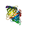
| ||||||||
|---|---|---|---|---|---|---|---|---|---|
| 1 |
| ||||||||
| Unit cell |
|
-Components
-Protein , 1 types, 1 molecules A
| #1: Protein | Mass: 22168.889 Da / Num. of mol.: 1 Source method: isolated from a genetically manipulated source Source: (gene. exp.) Ehrlichia chaffeensis str. Arkansas (bacteria)Gene: ECH_1053 / Plasmid: EhchA.17554.a.VL2 / Production host: |
|---|
-Non-polymers , 5 types, 10 molecules 


| #2: Chemical | ChemComp-SO4 /  Mass: 96.063 Da / Num. of mol.: 1 / Source method: obtained synthetically / Formula: SO4 Mass: 96.063 Da / Num. of mol.: 1 / Source method: obtained synthetically / Formula: SO4 |
|---|---|
| #3: Chemical | ChemComp-CL /  Mass: 35.453 Da / Num. of mol.: 1 / Source method: obtained synthetically / Formula: Cl Mass: 35.453 Da / Num. of mol.: 1 / Source method: obtained synthetically / Formula: Cl |
| #4: Chemical | ChemComp-GOL /  Mass: 92.094 Da / Num. of mol.: 1 / Source method: obtained synthetically / Formula: C3H8O3 Mass: 92.094 Da / Num. of mol.: 1 / Source method: obtained synthetically / Formula: C3H8O3 |
| #5: Chemical | ChemComp-PG4 /  Mass: 194.226 Da / Num. of mol.: 1 / Source method: obtained synthetically / Formula: C8H18O5 / Comment: precipitant*YM Mass: 194.226 Da / Num. of mol.: 1 / Source method: obtained synthetically / Formula: C8H18O5 / Comment: precipitant*YM |
| #6: Water | ChemComp-HOH / Mass: 18.015 Da / Num. of mol.: 6 / Source method: isolated from a natural source / Formula: H2O |
-Details
| Has ligand of interest | N |
|---|
-Experimental details
-Experiment
| Experiment | Method: X-RAY DIFFRACTION / Number of used crystals: 1 |
|---|
- Sample preparation
| Crystal | Density Matthews: 4.23 Å3/Da / Density % sol: 70.89 % |
|---|---|
| Crystal grow | Temperature: 291 K / Method: vapor diffusion, sitting drop / pH: 7 Details: Index HT C11 + 10% Hampton Additive E5: 1 M ammonium sulfate, 100 mM Hepes pH 7.0, 0.5% (w/v) PEG 8000, 1% (w/v) PEG 3350, EhchA.17554.a.VL2.PK00002 at 3.7 mg/mL. plate 13803 well E5 drop 1. ...Details: Index HT C11 + 10% Hampton Additive E5: 1 M ammonium sulfate, 100 mM Hepes pH 7.0, 0.5% (w/v) PEG 8000, 1% (w/v) PEG 3350, EhchA.17554.a.VL2.PK00002 at 3.7 mg/mL. plate 13803 well E5 drop 1. Puck: PSL-0312, Cryo: 80% crystallant + 20% glycerol. |
-Data collection
| Diffraction | Mean temperature: 100 K / Serial crystal experiment: N |
|---|---|
| Diffraction source | Source: SYNCHROTRON / Site: NSLS-II / Beamline: 19-ID / Wavelength: 0.9786 Å |
| Detector | Type: DECTRIS EIGER2 XE 9M / Detector: PIXEL / Date: Feb 14, 2024 |
| Radiation | Monochromator: Double Crystal Si 111 / Protocol: SINGLE WAVELENGTH / Monochromatic (M) / Laue (L): M / Scattering type: x-ray |
| Radiation wavelength | Wavelength: 0.9786 Å / Relative weight: 1 |
| Reflection | Resolution: 3→43.72 Å / Num. obs: 7533 / % possible obs: 100 % / Redundancy: 9.7 % / CC1/2: 0.994 / Rmerge(I) obs: 0.192 / Rpim(I) all: 0.066 / Rrim(I) all: 0.204 / Χ2: 1.01 / Net I/σ(I): 8.7 / Num. measured all: 73361 |
| Reflection shell | Resolution: 3→3.18 Å / % possible obs: 100 % / Redundancy: 10.8 % / Rmerge(I) obs: 1.317 / Num. measured all: 13177 / Num. unique obs: 1218 / CC1/2: 0.702 / Rpim(I) all: 0.419 / Rrim(I) all: 1.383 / Χ2: 0.94 / Net I/σ(I) obs: 2 |
- Processing
| Software |
| |||||||||||||||||||||||||||||||||||||||||||||||||||||||||||||||||||||||||||
|---|---|---|---|---|---|---|---|---|---|---|---|---|---|---|---|---|---|---|---|---|---|---|---|---|---|---|---|---|---|---|---|---|---|---|---|---|---|---|---|---|---|---|---|---|---|---|---|---|---|---|---|---|---|---|---|---|---|---|---|---|---|---|---|---|---|---|---|---|---|---|---|---|---|---|---|---|
| Refinement | Method to determine structure: MOLECULAR REPLACEMENT / Resolution: 3→43.72 Å / SU ML: 0.42 / Cross valid method: FREE R-VALUE / σ(F): 1.35 / Phase error: 30 / Stereochemistry target values: ML
| |||||||||||||||||||||||||||||||||||||||||||||||||||||||||||||||||||||||||||
| Solvent computation | Shrinkage radii: 0.9 Å / VDW probe radii: 1.1 Å / Solvent model: FLAT BULK SOLVENT MODEL | |||||||||||||||||||||||||||||||||||||||||||||||||||||||||||||||||||||||||||
| Refinement step | Cycle: LAST / Resolution: 3→43.72 Å
| |||||||||||||||||||||||||||||||||||||||||||||||||||||||||||||||||||||||||||
| Refine LS restraints |
| |||||||||||||||||||||||||||||||||||||||||||||||||||||||||||||||||||||||||||
| LS refinement shell |
| |||||||||||||||||||||||||||||||||||||||||||||||||||||||||||||||||||||||||||
| Refinement TLS params. | Method: refined / Refine-ID: X-RAY DIFFRACTION
| |||||||||||||||||||||||||||||||||||||||||||||||||||||||||||||||||||||||||||
| Refinement TLS group |
|
