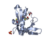
[English] 日本語
Yorodumi
- PDB-9c66: Structure of the Mena EVH1 domain bound to the polyproline segmen... -

+
Open data


ID or keywords:

Loading...

-
Basic information

Entry
Database: PDB / ID: 9c66
TitleStructure of the Mena EVH1 domain bound to the polyproline segment of PTP1B
Components
  • Protein enabled homolog
  • poly-proline segment of PTP1B
KeywordsSTRUCTURAL PROTEIN / Mena
Function / homology
Function and homology information


actin polymerization-dependent cell motility / profilin binding / Signaling by ROBO receptors / actin polymerization or depolymerization / WW domain binding / PTK6 Down-Regulation / regulation of hepatocyte growth factor receptor signaling pathway / positive regulation of receptor catabolic process / peptidyl-tyrosine dephosphorylation involved in inactivation of protein kinase activity / insulin receptor recycling ...actin polymerization-dependent cell motility / profilin binding / Signaling by ROBO receptors / actin polymerization or depolymerization / WW domain binding / PTK6 Down-Regulation / regulation of hepatocyte growth factor receptor signaling pathway / positive regulation of receptor catabolic process / peptidyl-tyrosine dephosphorylation involved in inactivation of protein kinase activity / insulin receptor recycling / negative regulation of vascular endothelial growth factor receptor signaling pathway / negative regulation of PERK-mediated unfolded protein response / regulation of intracellular protein transport / IRE1-mediated unfolded protein response / cytoplasmic side of endoplasmic reticulum membrane / sorting endosome / mitochondrial crista / platelet-derived growth factor receptor-beta signaling pathway / positive regulation of IRE1-mediated unfolded protein response / regulation of type I interferon-mediated signaling pathway / regulation of endocytosis / non-membrane spanning protein tyrosine phosphatase activity / positive regulation of protein tyrosine kinase activity / Generation of second messenger molecules / peptidyl-tyrosine dephosphorylation / Regulation of IFNA/IFNB signaling / regulation of signal transduction / cellular response to unfolded protein / growth hormone receptor signaling pathway via JAK-STAT / negative regulation of endoplasmic reticulum stress-induced intrinsic apoptotic signaling pathway / negative regulation of signal transduction / Regulation of IFNG signaling / Growth hormone receptor signaling / MECP2 regulates neuronal receptors and channels / endoplasmic reticulum unfolded protein response / negative regulation of MAP kinase activity / positive regulation of JUN kinase activity / negative regulation of insulin receptor signaling pathway / Insulin receptor recycling / ephrin receptor binding / Integrin signaling / protein dephosphorylation / protein-tyrosine-phosphatase / filopodium / protein tyrosine phosphatase activity / protein phosphatase 2A binding / axon guidance / endosome lumen / insulin receptor binding / Negative regulation of MET activity / receptor tyrosine kinase binding / negative regulation of ERK1 and ERK2 cascade / SH3 domain binding / cell junction / lamellipodium / insulin receptor signaling pathway / actin binding / actin cytoskeleton organization / early endosome / cytoskeleton / mitochondrial matrix / cadherin binding / focal adhesion / synapse / protein kinase binding / enzyme binding / endoplasmic reticulum / protein-containing complex / RNA binding / zinc ion binding / plasma membrane / cytosol / cytoplasm
Similarity search - Function
VASP tetramerisation / VASP tetramerisation domain superfamily / VASP tetramerisation domain / WH1/EVH1 domain / WH1 domain / WH1 domain profile. / WASP homology region 1 / Protein-tyrosine phosphatase, non-receptor type-1/2 / : / Protein tyrosine phosphatase, catalytic domain ...VASP tetramerisation / VASP tetramerisation domain superfamily / VASP tetramerisation domain / WH1/EVH1 domain / WH1 domain / WH1 domain profile. / WASP homology region 1 / Protein-tyrosine phosphatase, non-receptor type-1/2 / : / Protein tyrosine phosphatase, catalytic domain / PTP type protein phosphatase domain profile. / Tyrosine-specific protein phosphatase, PTPase domain / Protein-tyrosine phosphatase / Protein-tyrosine phosphatase, catalytic / Protein tyrosine phosphatase, catalytic domain motif / Tyrosine specific protein phosphatases active site. / Protein-tyrosine phosphatase, active site / Tyrosine-specific protein phosphatases domain / Tyrosine specific protein phosphatases domain profile. / Protein-tyrosine phosphatase-like / PH-like domain superfamily
Similarity search - Domain/homology
Tyrosine-protein phosphatase non-receptor type 1 / Protein enabled homolog
Similarity search - Component
Biological speciesHomo sapiens (human)
MethodX-RAY DIFFRACTION / SYNCHROTRON / MOLECULAR REPLACEMENT / Resolution: 1.4 Å
AuthorsLaComb, L. / Fedorov, E. / Bonanno, J.B. / Almo, S.C. / Ghosh, A.
Funding support United States, 1items
OrganizationGrant numberCountry
National Institutes of Health/National Cancer Institute (NIH/NCI) United States
CitationJournal: Biochemistry / Year: 2024
Title: Insights into the Interaction Landscape of the EVH1 Domain of Mena.
Authors: LaComb, L. / Ghosh, A. / Bonanno, J.B. / Nilson, D.J. / Poppel, A.J. / Dada, L. / Cahill, S.M. / Maianti, J.P. / Kitamura, S. / Cowburn, D. / Almo, S.C.
History
DepositionJun 7, 2024Deposition site: RCSB / Processing site: RCSB
Revision 1.0Aug 28, 2024Provider: repository / Type: Initial release
Revision 1.1Sep 11, 2024Group: Database references / Category: citation
Item: _citation.journal_volume / _citation.page_first / _citation.page_last

-
Structure visualization

Structure viewerMolecule:
MolmilJmol/JSmol

Downloads & links

-
Assembly

Deposited unit
A: Protein enabled homolog
B: poly-proline segment of PTP1B
hetero molecules


Theoretical massNumber of molelcules
Total (without water)14,2235
Polymers14,0032
Non-polymers2203
Water1,13563
1
A: Protein enabled homolog
hetero molecules


Theoretical massNumber of molelcules
Total (without water)13,0854
Polymers12,8651
Non-polymers2203
Water181
TypeNameSymmetry operationNumber
identity operation1_555x,y,z1
2
B: poly-proline segment of PTP1B


  • defined by author
  • 1.14 kDa, 1 polymers
Theoretical massNumber of molelcules
Total (without water)1,1381
Polymers1,1381
Non-polymers00
Water181
TypeNameSymmetry operationNumber
identity operation1_555x,y,z1
Unit cell
Length a, b, c (Å)39.654, 49.072, 54.105
Angle α, β, γ (deg.)90.00, 90.00, 90.00
Int Tables number19
Space group name H-MP212121

-
Components

#1: Protein Protein enabled homolog


Mass: 12864.520 Da / Num. of mol.: 1
Source method: isolated from a genetically manipulated source
Source: (gene. exp.) Homo sapiens (human) / Gene: ENAH, MENA / Production host: Escherichia coli BL21(DE3) (bacteria) / References: UniProt: Q8N8S7
#2: Protein/peptide poly-proline segment of PTP1B / Protein-tyrosine phosphatase 1B / PTP-1B


Mass: 1138.316 Da / Num. of mol.: 1 / Source method: obtained synthetically / Source: (synth.) Homo sapiens (human) / References: UniProt: P18031
#3: Chemical ChemComp-EDO / 1,2-ETHANEDIOL / ETHYLENE GLYCOL


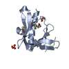
Mass: 62.068 Da / Num. of mol.: 2 / Source method: obtained synthetically / Formula: C2H6O2
#4: Chemical ChemComp-SO4 / SULFATE ION


Mass: 96.063 Da / Num. of mol.: 1 / Source method: obtained synthetically / Formula: SO4
#5: Water ChemComp-HOH / water


Mass: 18.015 Da / Num. of mol.: 63 / Source method: isolated from a natural source / Formula: H2O
Has ligand of interestN

-
Experimental details

-
Experiment

ExperimentMethod: X-RAY DIFFRACTION / Number of used crystals: 1

-
Sample preparation

CrystalDensity Matthews: 1.89 Å3/Da / Density % sol: 34.99 %
Crystal growTemperature: 292 K / Method: vapor diffusion, hanging drop / pH: 5.5 / Details: 2.0 M Ammonium sulfate and 0.1 M Bis-Tris

-
Data collection

DiffractionMean temperature: 100 K / Serial crystal experiment: N
Diffraction sourceSource: SYNCHROTRON / Site: APS / Beamline: 31-ID / Wavelength: 0.97932 Å
DetectorType: DECTRIS PILATUS 6M / Detector: PIXEL / Date: Jul 6, 2020
RadiationProtocol: SINGLE WAVELENGTH / Monochromatic (M) / Laue (L): M / Scattering type: x-ray
Radiation wavelengthWavelength: 0.97932 Å / Relative weight: 1
ReflectionResolution: 1.4→19.83 Å / Num. obs: 21372 / % possible obs: 99.6 % / Redundancy: 12.1 % / CC1/2: 0.998 / Rmerge(I) obs: 0.095 / Rpim(I) all: 0.029 / Rrim(I) all: 0.1 / Χ2: 1.01 / Net I/σ(I): 18.8 / Num. measured all: 259048
Reflection shellResolution: 1.4→1.42 Å / % possible obs: 100 % / Redundancy: 12 % / Rmerge(I) obs: 0.577 / Num. measured all: 12450 / Num. unique obs: 1040 / CC1/2: 0.889 / Rpim(I) all: 0.18 / Rrim(I) all: 0.606 / Χ2: 1.04 / Net I/σ(I) obs: 6.3

-
Processing

Software
NameVersionClassification
PHENIX(1.21rc1_5156)refinement
Aimlessdata scaling
XDSdata reduction
PHASERphasing
RefinementMethod to determine structure: MOLECULAR REPLACEMENT / Resolution: 1.4→19.83 Å / SU ML: 0.14 / Cross valid method: FREE R-VALUE / σ(F): 1.35 / Phase error: 20.05 / Stereochemistry target values: ML
RfactorNum. reflection% reflection
Rfree0.1961 1060 4.97 %
Rwork0.1873 --
obs0.1877 21323 99.63 %
Solvent computationShrinkage radii: 0.9 Å / VDW probe radii: 1.1 Å / Solvent model: FLAT BULK SOLVENT MODEL
Refinement stepCycle: LAST / Resolution: 1.4→19.83 Å
ProteinNucleic acidLigandSolventTotal
Num. atoms902 0 13 63 978
Refine LS restraints
Refine-IDTypeDev idealNumber
X-RAY DIFFRACTIONf_bond_d0.015
X-RAY DIFFRACTIONf_angle_d1.472
X-RAY DIFFRACTIONf_dihedral_angle_d16.943353
X-RAY DIFFRACTIONf_chiral_restr0.098146
X-RAY DIFFRACTIONf_plane_restr0.013177
LS refinement shell
Resolution (Å)Rfactor RfreeNum. reflection RfreeRfactor RworkNum. reflection RworkRefine-ID% reflection obs (%)
1.4-1.460.29261580.25212468X-RAY DIFFRACTION100
1.46-1.540.23161370.21622456X-RAY DIFFRACTION98
1.54-1.640.20581250.20092496X-RAY DIFFRACTION100
1.64-1.760.23131150.19042531X-RAY DIFFRACTION100
1.76-1.940.19071380.1792486X-RAY DIFFRACTION99
1.94-2.220.20111230.17352556X-RAY DIFFRACTION100
2.22-2.80.18581490.18132554X-RAY DIFFRACTION100
2.8-19.830.17161150.18352716X-RAY DIFFRACTION100

+
About Yorodumi

-
News

-
Feb 9, 2022. New format data for meta-information of EMDB entries

New format data for meta-information of EMDB entries

  • Version 3 of the EMDB header file is now the official format.
  • The previous official version 1.9 will be removed from the archive.

Related info.:EMDB header

External links:wwPDB to switch to version 3 of the EMDB data model

-
Aug 12, 2020. Covid-19 info

Covid-19 info

URL: https://pdbjlvh1.pdbj.org/emnavi/covid19.php

New page: Covid-19 featured information page in EM Navigator.

Related info.:Covid-19 info / Mar 5, 2020. Novel coronavirus structure data

+
Mar 5, 2020. Novel coronavirus structure data

Novel coronavirus structure data

Related info.:Yorodumi Speices / Aug 12, 2020. Covid-19 info

External links:COVID-19 featured content - PDBj / Molecule of the Month (242):Coronavirus Proteases

+
Jan 31, 2019. EMDB accession codes are about to change! (news from PDBe EMDB page)

EMDB accession codes are about to change! (news from PDBe EMDB page)

  • The allocation of 4 digits for EMDB accession codes will soon come to an end. Whilst these codes will remain in use, new EMDB accession codes will include an additional digit and will expand incrementally as the available range of codes is exhausted. The current 4-digit format prefixed with “EMD-” (i.e. EMD-XXXX) will advance to a 5-digit format (i.e. EMD-XXXXX), and so on. It is currently estimated that the 4-digit codes will be depleted around Spring 2019, at which point the 5-digit format will come into force.
  • The EM Navigator/Yorodumi systems omit the EMD- prefix.

Related info.:Q: What is EMD? / ID/Accession-code notation in Yorodumi/EM Navigator

External links:EMDB Accession Codes are Changing Soon! / Contact to PDBj

+
Jul 12, 2017. Major update of PDB

Major update of PDB

  • wwPDB released updated PDB data conforming to the new PDBx/mmCIF dictionary.
  • This is a major update changing the version number from 4 to 5, and with Remediation, in which all the entries are updated.
  • In this update, many items about electron microscopy experimental information are reorganized (e.g. em_software).
  • Now, EM Navigator and Yorodumi are based on the updated data.

External links:wwPDB Remediation / Enriched Model Files Conforming to OneDep Data Standards Now Available in the PDB FTP Archive

-
Yorodumi

Thousand views of thousand structures

  • Yorodumi is a browser for structure data from EMDB, PDB, SASBDB, etc.
  • This page is also the successor to EM Navigator detail page, and also detail information page/front-end page for Omokage search.
  • The word "yorodu" (or yorozu) is an old Japanese word meaning "ten thousand". "mi" (miru) is to see.

Related info.:EMDB / PDB / SASBDB / Comparison of 3 databanks / Yorodumi Search / Aug 31, 2016. New EM Navigator & Yorodumi / Yorodumi Papers / Jmol/JSmol / Function and homology information / Changes in new EM Navigator and Yorodumi

Read more