[English] 日本語
Yorodumi
- PDB-9c5s: Disulfide-linked, antiparallel p53-derived peptide dimer (CV1) -

+
Open data


ID or keywords:

Loading...

-
Basic information

Entry
Database: PDB / ID: 9c5s
TitleDisulfide-linked, antiparallel p53-derived peptide dimer (CV1)
ComponentsCellular tumor antigen p53
KeywordsUNKNOWN FUNCTION / Antiparallel dimer / p53
Function / homology
Function and homology information


Loss of function of TP53 in cancer due to loss of tetramerization ability / Regulation of TP53 Expression / negative regulation of helicase activity / signal transduction by p53 class mediator / negative regulation of G1 to G0 transition / negative regulation of glucose catabolic process to lactate via pyruvate / Transcriptional activation of cell cycle inhibitor p21 / regulation of intrinsic apoptotic signaling pathway by p53 class mediator / negative regulation of pentose-phosphate shunt / ATP-dependent DNA/DNA annealing activity ...Loss of function of TP53 in cancer due to loss of tetramerization ability / Regulation of TP53 Expression / negative regulation of helicase activity / signal transduction by p53 class mediator / negative regulation of G1 to G0 transition / negative regulation of glucose catabolic process to lactate via pyruvate / Transcriptional activation of cell cycle inhibitor p21 / regulation of intrinsic apoptotic signaling pathway by p53 class mediator / negative regulation of pentose-phosphate shunt / ATP-dependent DNA/DNA annealing activity / Activation of NOXA and translocation to mitochondria / regulation of cell cycle G2/M phase transition / regulation of fibroblast apoptotic process / intrinsic apoptotic signaling pathway in response to hypoxia / oligodendrocyte apoptotic process / negative regulation of miRNA processing / positive regulation of thymocyte apoptotic process / oxidative stress-induced premature senescence / regulation of tissue remodeling / glucose catabolic process to lactate via pyruvate / positive regulation of mitochondrial membrane permeability / positive regulation of programmed necrotic cell death / negative regulation of mitophagy / mRNA transcription / circadian behavior / bone marrow development / germ cell nucleus / regulation of mitochondrial membrane permeability involved in apoptotic process / regulation of DNA damage response, signal transduction by p53 class mediator / RUNX3 regulates CDKN1A transcription / TP53 regulates transcription of additional cell cycle genes whose exact role in the p53 pathway remain uncertain / TP53 Regulates Transcription of Death Receptors and Ligands / Activation of PUMA and translocation to mitochondria / : / histone deacetylase regulator activity / negative regulation of glial cell proliferation / Regulation of TP53 Activity through Association with Co-factors / negative regulation of neuroblast proliferation / T cell lineage commitment / mitochondrial DNA repair / Formation of Senescence-Associated Heterochromatin Foci (SAHF) / ER overload response / B cell lineage commitment / thymocyte apoptotic process / TP53 Regulates Transcription of Caspase Activators and Caspases / negative regulation of DNA replication / cardiac septum morphogenesis / entrainment of circadian clock by photoperiod / PI5P Regulates TP53 Acetylation / negative regulation of telomere maintenance via telomerase / necroptotic process / positive regulation of release of cytochrome c from mitochondria / Zygotic genome activation (ZGA) / Association of TriC/CCT with target proteins during biosynthesis / TP53 Regulates Transcription of Genes Involved in Cytochrome C Release / TFIID-class transcription factor complex binding / T cell proliferation involved in immune response / rRNA transcription / SUMOylation of transcription factors / TP53 regulates transcription of several additional cell death genes whose specific roles in p53-dependent apoptosis remain uncertain / intrinsic apoptotic signaling pathway by p53 class mediator / positive regulation of execution phase of apoptosis / Transcriptional Regulation by VENTX / general transcription initiation factor binding / replicative senescence / cellular response to actinomycin D / intrinsic apoptotic signaling pathway in response to endoplasmic reticulum stress / cellular response to UV-C / neuroblast proliferation / hematopoietic stem cell differentiation / positive regulation of RNA polymerase II transcription preinitiation complex assembly / response to X-ray / negative regulation of reactive oxygen species metabolic process / intrinsic apoptotic signaling pathway in response to DNA damage by p53 class mediator / DNA damage response, signal transduction by p53 class mediator / chromosome organization / viral process / Pyroptosis / embryonic organ development / cis-regulatory region sequence-specific DNA binding / type II interferon-mediated signaling pathway / TP53 Regulates Transcription of Genes Involved in G1 Cell Cycle Arrest / somitogenesis / glial cell proliferation / hematopoietic progenitor cell differentiation / core promoter sequence-specific DNA binding / negative regulation of stem cell proliferation / : / cellular response to glucose starvation / mitophagy / positive regulation of intrinsic apoptotic signaling pathway / negative regulation of fibroblast proliferation / positive regulation of cardiac muscle cell apoptotic process / tumor necrosis factor-mediated signaling pathway / mitotic G1 DNA damage checkpoint signaling / Regulation of TP53 Activity through Acetylation / gastrulation / MDM2/MDM4 family protein binding / response to salt stress / 14-3-3 protein binding
Similarity search - Function
Cellular tumor antigen p53, transactivation domain 2 / Transactivation domain 2 / p53 transactivation domain / P53 transactivation motif / p53 family signature. / p53, tetramerisation domain / P53 tetramerisation motif / p53, DNA-binding domain / P53 DNA-binding domain / p53 tumour suppressor family ...Cellular tumor antigen p53, transactivation domain 2 / Transactivation domain 2 / p53 transactivation domain / P53 transactivation motif / p53 family signature. / p53, tetramerisation domain / P53 tetramerisation motif / p53, DNA-binding domain / P53 DNA-binding domain / p53 tumour suppressor family / p53-like tetramerisation domain superfamily / p53/RUNT-type transcription factor, DNA-binding domain superfamily / p53-like transcription factor, DNA-binding
Similarity search - Domain/homology
Cellular tumor antigen p53
Similarity search - Component
Biological speciesHomo sapiens (human)
MethodX-RAY DIFFRACTION / SYNCHROTRON / AB INITIO PHASING / Resolution: 1.01 Å
AuthorsVithanage, N. / Kreitler, D.K. / DiGiorno, M.C. / Victorio, C.G. / Sawyer, N. / Outlaw, V.K.
Funding support1items
OrganizationGrant numberCountry
Not funded
CitationJournal: Res Sq / Year: 2024
Title: Structural Characterization of Disulfide-Linked p53-Derived Peptide Dimers.
Authors: DiGiorno, M.C. / Vithanage, N. / Victorio, C.G. / Kreitler, D.F. / Outlaw, V.K. / Sawyer, N.
History
DepositionJun 6, 2024Deposition site: RCSB / Processing site: RCSB
Revision 1.0Jun 26, 2024Provider: repository / Type: Initial release
Revision 1.1Aug 7, 2024Group: Database references / Category: citation / citation_author
Item: _citation.country / _citation.journal_abbrev ..._citation.country / _citation.journal_abbrev / _citation.journal_id_CSD / _citation.journal_id_ISSN / _citation.pdbx_database_id_DOI / _citation.pdbx_database_id_PubMed / _citation.title / _citation.year / _citation_author.identifier_ORCID / _citation_author.name
Revision 1.2Oct 16, 2024Group: Structure summary / Category: pdbx_entry_details / pdbx_modification_feature / Item: _pdbx_entry_details.has_protein_modification

-
Structure visualization

Structure viewerMolecule:
MolmilJmol/JSmol

Downloads & links

-
Assembly

Deposited unit
A: Cellular tumor antigen p53
B: Cellular tumor antigen p53
C: Cellular tumor antigen p53
D: Cellular tumor antigen p53
hetero molecules


Theoretical massNumber of molelcules
Total (without water)6,8085
Polymers6,7124
Non-polymers961
Water1,27971
1


  • Idetical with deposited unit
  • defined by author&software
TypeNameSymmetry operationNumber
identity operation1_555x,y,z1
Buried area4200 Å2
ΔGint-63 kcal/mol
Surface area3320 Å2
MethodPISA
Unit cell
Length a, b, c (Å)45.243, 45.243, 62.699
Angle α, β, γ (deg.)90.000, 90.000, 120.000
Int Tables number152
Space group name H-MP3121
Space group name HallP312"
Symmetry operation#1: x,y,z
#2: -y,x-y,z+1/3
#3: -x+y,-x,z+2/3
#4: x-y,-y,-z+2/3
#5: -x,-x+y,-z+1/3
#6: y,x,-z

-
Components

#1: Protein/peptide
Cellular tumor antigen p53 / Antigen NY-CO-13 / Phosphoprotein p53 / Tumor suppressor p53


Mass: 1677.991 Da / Num. of mol.: 4 / Mutation: p53-derived / Source method: obtained synthetically
Details: This molecule is a synthetic peptide derived from residues 17-30 of the p53 protein. It is acetylated at the N-terminus and amidated at the C-terminus.
Source: (synth.) Homo sapiens (human) / References: UniProt: P04637
#2: Chemical ChemComp-SO4 / SULFATE ION


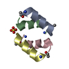
Mass: 96.063 Da / Num. of mol.: 1 / Source method: isolated from a natural source / Formula: SO4
#3: Water ChemComp-HOH / water


Mass: 18.015 Da / Num. of mol.: 71 / Source method: isolated from a natural source / Formula: H2O
Has ligand of interestN
Has protein modificationY

-
Experimental details

-
Experiment

ExperimentMethod: X-RAY DIFFRACTION / Number of used crystals: 1

-
Sample preparation

CrystalDensity Matthews: 2.76 Å3/Da / Density % sol: 55.42 %
Crystal growTemperature: 291 K / Method: vapor diffusion, hanging drop / pH: 6.5
Details: 30 mM sodium nitrate, 30 mM dibasic sodium phosphate, 30 mM ammonium sulfate, 20% v/v glycerol, 10% w/v PEG4000, 100 mM imidazole/MES monohydrate buffer, pH 6.5

-
Data collection

DiffractionMean temperature: 100 K / Serial crystal experiment: N
Diffraction sourceSource: SYNCHROTRON / Site: NSLS-II / Beamline: 17-ID-1 / Wavelength: 0.92011 Å
DetectorType: DECTRIS EIGER X 9M / Detector: PIXEL / Date: Sep 17, 2023
RadiationProtocol: SINGLE WAVELENGTH / Monochromatic (M) / Laue (L): M / Scattering type: x-ray
Radiation wavelengthWavelength: 0.92011 Å / Relative weight: 1
ReflectionResolution: 1.01→33.23 Å / Num. obs: 35044 / % possible obs: 88.6 % / Redundancy: 10.6 % / Biso Wilson estimate: 14.12 Å2 / CC1/2: 0.999 / Rmerge(I) obs: 0.045 / Net I/σ(I): 15
Reflection shellResolution: 1.01→1.068 Å / Redundancy: 7.6 % / Rmerge(I) obs: 1.553 / Num. unique obs: 1752 / CC1/2: 0.524 / % possible all: 28.9

-
Processing

Software
NameVersionClassification
PHENIX1.20.1_4487refinement
PHASERphasing
XDSdata reduction
Aimlessdata scaling
RefinementMethod to determine structure: AB INITIO PHASING / Resolution: 1.01→33.23 Å / SU ML: 0.1037 / Cross valid method: FREE R-VALUE / σ(F): 1.34 / Phase error: 29.9692
Stereochemistry target values: GeoStd + Monomer Library + CDL v1.2
RfactorNum. reflection% reflection
Rfree0.2015 1699 4.85 %
Rwork0.1904 33342 -
obs0.1909 35041 88.56 %
Solvent computationShrinkage radii: 0.9 Å / VDW probe radii: 1.1 Å / Solvent model: FLAT BULK SOLVENT MODEL
Displacement parametersBiso mean: 21.87 Å2
Refinement stepCycle: LAST / Resolution: 1.01→33.23 Å
ProteinNucleic acidLigandSolventTotal
Num. atoms472 0 5 71 548
Refine LS restraints
Refine-IDTypeDev idealNumber
X-RAY DIFFRACTIONf_bond_d0.0044484
X-RAY DIFFRACTIONf_angle_d0.8244658
X-RAY DIFFRACTIONf_chiral_restr0.054172
X-RAY DIFFRACTIONf_plane_restr0.004984
X-RAY DIFFRACTIONf_dihedral_angle_d8.566164
LS refinement shell
Resolution (Å)Rfactor RfreeNum. reflection RfreeRfactor RworkNum. reflection RworkRefine-ID% reflection obs (%)
1.01-1.040.3494170.376305X-RAY DIFFRACTION10.01
1.04-1.070.28631000.31951698X-RAY DIFFRACTION55.14
1.07-1.110.28891350.27883029X-RAY DIFFRACTION97.5
1.11-1.160.24831210.26693142X-RAY DIFFRACTION100
1.16-1.210.21071500.23223117X-RAY DIFFRACTION99.97
1.21-1.270.26821420.21823118X-RAY DIFFRACTION100
1.27-1.350.22661790.20563097X-RAY DIFFRACTION100
1.35-1.460.22181540.19923155X-RAY DIFFRACTION99.97
1.46-1.60.1751730.18953109X-RAY DIFFRACTION100
1.6-1.830.1861590.18063151X-RAY DIFFRACTION100
1.83-2.310.1621860.17723169X-RAY DIFFRACTION100
2.31-33.230.21391830.18153252X-RAY DIFFRACTION98.23
Refinement TLS params.Method: refined / Origin x: -13.911 Å / Origin y: 10.57 Å / Origin z: 4.425 Å
111213212223313233
T0.126795389039 Å2-0.0268009730784 Å2-0.00843600895083 Å2-0.101230355949 Å2-0.00389821042246 Å2--0.103016014259 Å2
L3.24313455436 °21.27315579524 °22.60762407649 °2-3.08974919546 °21.72461217933 °2--4.54394151365 °2
S0.0307888780188 Å °0.0147582035078 Å °-0.16560641241 Å °-0.026233619184 Å °0.104845157707 Å °-0.131982514352 Å °0.325824015357 Å °0.0287843624297 Å °-0.068269696598 Å °
Refinement TLS group
IDRefine-IDRefine TLS-IDSelection detailsAuth asym-IDAuth seq-ID
1X-RAY DIFFRACTION1( CHAIN A AND ( RESID 0:14 OR RESID 15:15 OR RESID 101:118 ) ) OR ( CHAIN B AND ( RESID 0:14 OR RESID 15:15 OR RESID 201:225 OR RESID 101:101 ) ) OR ( CHAIN C AND ( RESID 0:14 OR RESID 15:15 OR RESID 101:111 ) ) OR ( CHAIN D AND ( RESID 0:14 OR RESID 15:15 OR RESID 101:117 ) )A0 - 14
2X-RAY DIFFRACTION1( CHAIN A AND ( RESID 0:14 OR RESID 15:15 OR RESID 101:118 ) ) OR ( CHAIN B AND ( RESID 0:14 OR RESID 15:15 OR RESID 201:225 OR RESID 101:101 ) ) OR ( CHAIN C AND ( RESID 0:14 OR RESID 15:15 OR RESID 101:111 ) ) OR ( CHAIN D AND ( RESID 0:14 OR RESID 15:15 OR RESID 101:117 ) )A15
3X-RAY DIFFRACTION1( CHAIN A AND ( RESID 0:14 OR RESID 15:15 OR RESID 101:118 ) ) OR ( CHAIN B AND ( RESID 0:14 OR RESID 15:15 OR RESID 201:225 OR RESID 101:101 ) ) OR ( CHAIN C AND ( RESID 0:14 OR RESID 15:15 OR RESID 101:111 ) ) OR ( CHAIN D AND ( RESID 0:14 OR RESID 15:15 OR RESID 101:117 ) )A101 - 118
4X-RAY DIFFRACTION1( CHAIN A AND ( RESID 0:14 OR RESID 15:15 OR RESID 101:118 ) ) OR ( CHAIN B AND ( RESID 0:14 OR RESID 15:15 OR RESID 201:225 OR RESID 101:101 ) ) OR ( CHAIN C AND ( RESID 0:14 OR RESID 15:15 OR RESID 101:111 ) ) OR ( CHAIN D AND ( RESID 0:14 OR RESID 15:15 OR RESID 101:117 ) )B0 - 14
5X-RAY DIFFRACTION1( CHAIN A AND ( RESID 0:14 OR RESID 15:15 OR RESID 101:118 ) ) OR ( CHAIN B AND ( RESID 0:14 OR RESID 15:15 OR RESID 201:225 OR RESID 101:101 ) ) OR ( CHAIN C AND ( RESID 0:14 OR RESID 15:15 OR RESID 101:111 ) ) OR ( CHAIN D AND ( RESID 0:14 OR RESID 15:15 OR RESID 101:117 ) )B15
6X-RAY DIFFRACTION1( CHAIN A AND ( RESID 0:14 OR RESID 15:15 OR RESID 101:118 ) ) OR ( CHAIN B AND ( RESID 0:14 OR RESID 15:15 OR RESID 201:225 OR RESID 101:101 ) ) OR ( CHAIN C AND ( RESID 0:14 OR RESID 15:15 OR RESID 101:111 ) ) OR ( CHAIN D AND ( RESID 0:14 OR RESID 15:15 OR RESID 101:117 ) )B201 - 225
7X-RAY DIFFRACTION1( CHAIN A AND ( RESID 0:14 OR RESID 15:15 OR RESID 101:118 ) ) OR ( CHAIN B AND ( RESID 0:14 OR RESID 15:15 OR RESID 201:225 OR RESID 101:101 ) ) OR ( CHAIN C AND ( RESID 0:14 OR RESID 15:15 OR RESID 101:111 ) ) OR ( CHAIN D AND ( RESID 0:14 OR RESID 15:15 OR RESID 101:117 ) )B101
8X-RAY DIFFRACTION1( CHAIN A AND ( RESID 0:14 OR RESID 15:15 OR RESID 101:118 ) ) OR ( CHAIN B AND ( RESID 0:14 OR RESID 15:15 OR RESID 201:225 OR RESID 101:101 ) ) OR ( CHAIN C AND ( RESID 0:14 OR RESID 15:15 OR RESID 101:111 ) ) OR ( CHAIN D AND ( RESID 0:14 OR RESID 15:15 OR RESID 101:117 ) )C0 - 14
9X-RAY DIFFRACTION1( CHAIN A AND ( RESID 0:14 OR RESID 15:15 OR RESID 101:118 ) ) OR ( CHAIN B AND ( RESID 0:14 OR RESID 15:15 OR RESID 201:225 OR RESID 101:101 ) ) OR ( CHAIN C AND ( RESID 0:14 OR RESID 15:15 OR RESID 101:111 ) ) OR ( CHAIN D AND ( RESID 0:14 OR RESID 15:15 OR RESID 101:117 ) )C15
10X-RAY DIFFRACTION1( CHAIN A AND ( RESID 0:14 OR RESID 15:15 OR RESID 101:118 ) ) OR ( CHAIN B AND ( RESID 0:14 OR RESID 15:15 OR RESID 201:225 OR RESID 101:101 ) ) OR ( CHAIN C AND ( RESID 0:14 OR RESID 15:15 OR RESID 101:111 ) ) OR ( CHAIN D AND ( RESID 0:14 OR RESID 15:15 OR RESID 101:117 ) )C101 - 111
11X-RAY DIFFRACTION1( CHAIN A AND ( RESID 0:14 OR RESID 15:15 OR RESID 101:118 ) ) OR ( CHAIN B AND ( RESID 0:14 OR RESID 15:15 OR RESID 201:225 OR RESID 101:101 ) ) OR ( CHAIN C AND ( RESID 0:14 OR RESID 15:15 OR RESID 101:111 ) ) OR ( CHAIN D AND ( RESID 0:14 OR RESID 15:15 OR RESID 101:117 ) )D0 - 14
12X-RAY DIFFRACTION1( CHAIN A AND ( RESID 0:14 OR RESID 15:15 OR RESID 101:118 ) ) OR ( CHAIN B AND ( RESID 0:14 OR RESID 15:15 OR RESID 201:225 OR RESID 101:101 ) ) OR ( CHAIN C AND ( RESID 0:14 OR RESID 15:15 OR RESID 101:111 ) ) OR ( CHAIN D AND ( RESID 0:14 OR RESID 15:15 OR RESID 101:117 ) )D15
13X-RAY DIFFRACTION1( CHAIN A AND ( RESID 0:14 OR RESID 15:15 OR RESID 101:118 ) ) OR ( CHAIN B AND ( RESID 0:14 OR RESID 15:15 OR RESID 201:225 OR RESID 101:101 ) ) OR ( CHAIN C AND ( RESID 0:14 OR RESID 15:15 OR RESID 101:111 ) ) OR ( CHAIN D AND ( RESID 0:14 OR RESID 15:15 OR RESID 101:117 ) )D101 - 117

+
About Yorodumi

-
News

-
Feb 9, 2022. New format data for meta-information of EMDB entries

New format data for meta-information of EMDB entries

  • Version 3 of the EMDB header file is now the official format.
  • The previous official version 1.9 will be removed from the archive.

Related info.:EMDB header

External links:wwPDB to switch to version 3 of the EMDB data model

-
Aug 12, 2020. Covid-19 info

Covid-19 info

URL: https://pdbj.org/emnavi/covid19.php

New page: Covid-19 featured information page in EM Navigator.

Related info.:Covid-19 info / Mar 5, 2020. Novel coronavirus structure data

+
Mar 5, 2020. Novel coronavirus structure data

Novel coronavirus structure data

Related info.:Yorodumi Speices / Aug 12, 2020. Covid-19 info

External links:COVID-19 featured content - PDBj / Molecule of the Month (242):Coronavirus Proteases

+
Jan 31, 2019. EMDB accession codes are about to change! (news from PDBe EMDB page)

EMDB accession codes are about to change! (news from PDBe EMDB page)

  • The allocation of 4 digits for EMDB accession codes will soon come to an end. Whilst these codes will remain in use, new EMDB accession codes will include an additional digit and will expand incrementally as the available range of codes is exhausted. The current 4-digit format prefixed with “EMD-” (i.e. EMD-XXXX) will advance to a 5-digit format (i.e. EMD-XXXXX), and so on. It is currently estimated that the 4-digit codes will be depleted around Spring 2019, at which point the 5-digit format will come into force.
  • The EM Navigator/Yorodumi systems omit the EMD- prefix.

Related info.:Q: What is EMD? / ID/Accession-code notation in Yorodumi/EM Navigator

External links:EMDB Accession Codes are Changing Soon! / Contact to PDBj

+
Jul 12, 2017. Major update of PDB

Major update of PDB

  • wwPDB released updated PDB data conforming to the new PDBx/mmCIF dictionary.
  • This is a major update changing the version number from 4 to 5, and with Remediation, in which all the entries are updated.
  • In this update, many items about electron microscopy experimental information are reorganized (e.g. em_software).
  • Now, EM Navigator and Yorodumi are based on the updated data.

External links:wwPDB Remediation / Enriched Model Files Conforming to OneDep Data Standards Now Available in the PDB FTP Archive

-
Yorodumi

Thousand views of thousand structures

  • Yorodumi is a browser for structure data from EMDB, PDB, SASBDB, etc.
  • This page is also the successor to EM Navigator detail page, and also detail information page/front-end page for Omokage search.
  • The word "yorodu" (or yorozu) is an old Japanese word meaning "ten thousand". "mi" (miru) is to see.

Related info.:EMDB / PDB / SASBDB / Comparison of 3 databanks / Yorodumi Search / Aug 31, 2016. New EM Navigator & Yorodumi / Yorodumi Papers / Jmol/JSmol / Function and homology information / Changes in new EM Navigator and Yorodumi

Read more