 Movie
Movie Controller

[English] 日本語
 Yorodumi
- PDB-9c1g: Rhesus rotavirus (consensus structure at 2.36 Angstrom resolution) -
+ Open data

- Basic information
| Entry | Database: PDB / ID: 9c1g | ||||||||||||||||||||||||||||||
|---|---|---|---|---|---|---|---|---|---|---|---|---|---|---|---|---|---|---|---|---|---|---|---|---|---|---|---|---|---|---|---|
| Title | Rhesus rotavirus (consensus structure at 2.36 Angstrom resolution) | ||||||||||||||||||||||||||||||
 Components Components |
| ||||||||||||||||||||||||||||||
 Keywords Keywords | VIRUS / Rotavirus / non-enveloped virus / viral particle | ||||||||||||||||||||||||||||||
| Function / homology |  Function and homology information Function and homology informationviral intermediate capsid / host cell endoplasmic reticulum lumen / T=13 icosahedral viral capsid / T=2 icosahedral viral capsid / viral inner capsid / viral outer capsid / viral nucleocapsid / receptor-mediated virion attachment to host cell / host cell surface receptor binding / fusion of virus membrane with host plasma membrane ...viral intermediate capsid / host cell endoplasmic reticulum lumen / T=13 icosahedral viral capsid / T=2 icosahedral viral capsid / viral inner capsid / viral outer capsid / viral nucleocapsid / receptor-mediated virion attachment to host cell / host cell surface receptor binding / fusion of virus membrane with host plasma membrane / viral envelope / structural molecule activity / RNA binding / metal ion binding Similarity search - Function | ||||||||||||||||||||||||||||||
| Biological species |  Simian rotavirus A strain RRV Simian rotavirus A strain RRV | ||||||||||||||||||||||||||||||
| Method | ELECTRON MICROSCOPY / single particle reconstruction / cryo EM / Resolution: 2.36 Å | ||||||||||||||||||||||||||||||
 Authors Authors | Jenni, S. / Herrmann, T. / De Sautu, M. / Harrison, S.C. | ||||||||||||||||||||||||||||||
| Funding support |  United States, 1items United States, 1items
| ||||||||||||||||||||||||||||||
 Citation Citation | Journal: To Be Published Title: Rotavirus structure Authors: Jenni, S. / Herrmann, T. / De Sautu, M. / Harrison, S.C. | ||||||||||||||||||||||||||||||
| History |
|
- Structure visualization
| Structure viewer | Molecule: MolmilJmol/JSmol |
|---|
- Downloads & links
-Download
| PDBx/mmCIF format | 9c1g.cif.gz | 4.5 MB | Display | PDBx/mmCIF format |
|---|---|---|---|---|
| PDB format | pdb9c1g.ent.gz | Display | PDB format | |
| PDBx/mmJSON format | 9c1g.json.gz | Tree view | PDBx/mmJSON format | |
| Others |  Other downloads Other downloads |
-Validation report
| Arichive directory | https://data.pdbj.org/pub/pdb/validation_reports/c1/9c1gftp://data.pdbj.org/pub/pdb/validation_reports/c1/9c1g | HTTPS FTP |
|---|
-Related structure data
| Related structure data |  45118MC 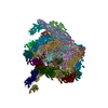 9c1hC  9c1iC  9c1jC  9c1kC  9c1lC M: map data used to model this data C: citing same article ( |
|---|---|
| Similar structure data |


-Links
 PDBj
PDBj


- Assembly
| Deposited unit | 
|
|---|---|
| 1 | x 60
|
| 2 |
|
| Symmetry | Point symmetry: (Schoenflies symbol: C1 (asymmetric)) |
-Components
-Protein , 3 types, 28 molecules 01PQRSTUVWXYZABCDEFGHIJKLMNO
| #1: Protein | Mass: 37136.531 Da / Num. of mol.: 13 Source method: isolated from a genetically manipulated source Source: (gene. exp.) Simian rotavirus A strain RRV / Production host: Simian rotavirus A strain RRV / References: UniProt: P12476#2: Protein | Mass: 103425.992 Da / Num. of mol.: 2 Source method: isolated from a genetically manipulated source Source: (gene. exp.) Simian rotavirus A strain RRV / Production host: Simian rotavirus A strain RRV / References: UniProt: B3F2X3#3: Protein | Mass: 44962.773 Da / Num. of mol.: 13 Source method: isolated from a genetically manipulated source Source: (gene. exp.) Simian rotavirus A strain RRV / Production host: Simian rotavirus A strain RRV / References: UniProt: B2BN53 |
|---|
-Sugars , 1 types, 13 molecules 
| #4: Sugar | ChemComp-NAG /  Type: D-saccharide, beta linking / Mass: 221.208 Da / Num. of mol.: 13 / Source method: obtained synthetically / Formula: C8H15NO6 Type: D-saccharide, beta linking / Mass: 221.208 Da / Num. of mol.: 13 / Source method: obtained synthetically / Formula: C8H15NO6 |
|---|
-Non-polymers , 3 types, 75 molecules 


| #5: Chemical | ChemComp-CA /  Mass: 40.078 Da / Num. of mol.: 52 / Source method: obtained synthetically / Formula: Ca Mass: 40.078 Da / Num. of mol.: 52 / Source method: obtained synthetically / Formula: Ca#6: Chemical | ChemComp-ZN /  Mass: 65.409 Da / Num. of mol.: 18 / Source method: obtained synthetically / Formula: Zn Mass: 65.409 Da / Num. of mol.: 18 / Source method: obtained synthetically / Formula: Zn#7: Chemical | ChemComp-CL /  Mass: 35.453 Da / Num. of mol.: 5 / Source method: obtained synthetically / Formula: Cl Mass: 35.453 Da / Num. of mol.: 5 / Source method: obtained synthetically / Formula: Cl |
|---|
-Details
| Has ligand of interest | N |
|---|---|
| Has protein modification | Y |
-Experimental details
-Experiment
| Experiment | Method: ELECTRON MICROSCOPY |
|---|---|
| EM experiment | Aggregation state: PARTICLE / 3D reconstruction method: single particle reconstruction |
- Sample preparation
| Component | Name: Simian rotavirus A strain RRV / Type: VIRUS / Entity ID: #1-#3 / Source: RECOMBINANT |
|---|---|
| Source (natural) | Organism: Simian rotavirus A strain RRV |
| Source (recombinant) | Organism: Simian rotavirus A strain RRV |
| Details of virus | Empty: NO / Enveloped: NO / Isolate: STRAIN / Type: VIRION |
| Buffer solution | pH: 8 |
| Specimen | Embedding applied: NO / Shadowing applied: NO / Staining applied: NO / Vitrification applied: YES |
| Vitrification | Cryogen name: ETHANE |
- Electron microscopy imaging
| Experimental equipment |  Model: Titan Krios / Image courtesy: FEI Company |
|---|---|
| Microscopy | Model: FEI TITAN KRIOS |
| Electron gun | Electron source:  FIELD EMISSION GUN / Accelerating voltage: 300 kV / Illumination mode: FLOOD BEAM FIELD EMISSION GUN / Accelerating voltage: 300 kV / Illumination mode: FLOOD BEAM |
| Electron lens | Mode: BRIGHT FIELD / Nominal defocus max: 2500 nm / Nominal defocus min: 1000 nm |
| Image recording | Electron dose: 50 e/Å2 / Film or detector model: GATAN K3 (6k x 4k) |
- Processing
| EM software | Name: PHENIX / Version: 1.21rc1_5127 / Category: model refinement | ||||||||||||||||||||||||
|---|---|---|---|---|---|---|---|---|---|---|---|---|---|---|---|---|---|---|---|---|---|---|---|---|---|
| CTF correction | Type: PHASE FLIPPING AND AMPLITUDE CORRECTION | ||||||||||||||||||||||||
| 3D reconstruction | Resolution: 2.36 Å / Resolution method: FSC 0.143 CUT-OFF / Num. of particles: 75846 / Symmetry type: POINT | ||||||||||||||||||||||||
| Refinement | Cross valid method: NONE Stereochemistry target values: GeoStd + Monomer Library + CDL v1.2 | ||||||||||||||||||||||||
| Displacement parameters | Biso mean: 24.91 Å2 | ||||||||||||||||||||||||
| Refine LS restraints |
|
