Entry Database : PDB / ID : 9c0yTitle Clathrin terminal domain complexed with Pitstop 2c Clathrin heavy chain 1 Keywords / Function / homology Function Domain/homology Component
/ / / / / / / / / / / / / / / / / / / / / / / / / / / / / / / / / / / / / / / / / / / / / / / / / / / / / / / / / / / / / / / / / / / / / / / / / / / / / / / / / / / / / / / / / / / / / / / / / Biological species Homo sapiens (human)Method / / / Resolution : 1.4 Å Authors Bulut, H. / Horatscheck, A. / Krauss, M. / Santos, K.F. / McCluskey, A. / Wahl, C.W. / Nazare, M. / Haucke, V. Funding support Organization Grant number Country Leibniz Association
Journal : Structure / Year : 2025Title : Next-generation small molecule inhibitors of clathrin function acutely inhibit endocytosis.Authors: Horatscheck, A. / Krauss, M. / Bulut, H. / Chambon, V. / Zadah, M.S. / Dransart, E. / Peloza, K. / Santos, K.F. / Robertson, M.J. / Prichard, K. / Miksche, S. / Radetzki, S. / von Kries, J.P. ... Authors : Horatscheck, A. / Krauss, M. / Bulut, H. / Chambon, V. / Zadah, M.S. / Dransart, E. / Peloza, K. / Santos, K.F. / Robertson, M.J. / Prichard, K. / Miksche, S. / Radetzki, S. / von Kries, J.P. / Wahl, M.C. / McCluskey, A. / Johannes, L. / Haucke, V. / Nazare, M. History Deposition May 28, 2024 Deposition site / Processing site Revision 1.0 Jun 26, 2024 Provider / Type Revision 1.1 Apr 16, 2025 Group / Structure summary / Category / citation_author / pdbx_entry_detailsItem _citation.country / _citation.journal_abbrev ... _citation.country / _citation.journal_abbrev / _citation.journal_id_ASTM / _citation.journal_id_CSD / _citation.journal_id_ISSN / _citation.pdbx_database_id_DOI / _citation.pdbx_database_id_PubMed / _citation.title / _citation.year / _pdbx_entry_details.has_protein_modification Revision 1.2 May 14, 2025 Group / Category / Item / _citation.page_first
Show all Show less
 Movie
Movie Controller


 Open data
 Basic information
 Components
 Keywords
 Function and homology information
 Homo sapiens (human)
 X-RAY DIFFRACTION /
  Authors
 Germany, 1items
  Citation
 Structure visualization
 Downloads & links
 Other downloads
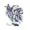
 PDBj
PDBj


 Assembly


 Mass: 78.133 Da / Num. of mol.: 2 / Source method: obtained synthetically / Formula: C2H6OS / Comment: DMSO, precipitant*YM
 Mass: 106.120 Da / Num. of mol.: 1 / Source method: obtained synthetically / Formula: C4H10O3
 Mass: 62.068 Da / Num. of mol.: 4 / Source method: obtained synthetically / Formula: C2H6O2
 Mass: 92.094 Da / Num. of mol.: 3 / Source method: obtained synthetically / Formula: C3H8O3
 Mass: 59.044 Da / Num. of mol.: 2 / Source method: obtained synthetically / Formula: C2H3O2
 Sample preparation
 Processing