 Movie
Movie Controller

[English] 日本語
 Yorodumi
- PDB-9bvm: Vitamin K-dependent gamma-carboxylase with protein C propeptide a... -
+ Open data

- Basic information
| Entry | Database: PDB / ID: 9bvm | ||||||||||||||||||||||||
|---|---|---|---|---|---|---|---|---|---|---|---|---|---|---|---|---|---|---|---|---|---|---|---|---|---|
| Title | Vitamin K-dependent gamma-carboxylase with protein C propeptide and glutamate-rich region and with vitamin K hydroquinone | ||||||||||||||||||||||||
 Components Components |
| ||||||||||||||||||||||||
 Keywords Keywords | MEMBRANE PROTEIN / LYASE/SUBSTRATE / GGCX / VKGC / Vitamin K / VKCFD / Hemophilia B / Warfarin / Carboxylation / Blood Coagulaton / Calcium homeostasis / Protein C / LYASE-SUBSTRATE complex | ||||||||||||||||||||||||
| Function / homology |  Function and homology information Function and homology informationactivated protein C (thrombin-activated peptidase) / positive regulation of establishment of endothelial barrier / peptidyl-glutamate 4-carboxylase / gamma-glutamyl carboxylase activity / negative regulation of coagulation / negative regulation of testosterone biosynthetic process / negative regulation of bone development / Defective gamma-carboxylation of F9 / vitamin binding / vitamin K metabolic process ...activated protein C (thrombin-activated peptidase) / positive regulation of establishment of endothelial barrier / peptidyl-glutamate 4-carboxylase / gamma-glutamyl carboxylase activity / negative regulation of coagulation / negative regulation of testosterone biosynthetic process / negative regulation of bone development / Defective gamma-carboxylation of F9 / vitamin binding / vitamin K metabolic process / negative regulation of neurotransmitter secretion / type B pancreatic cell proliferation / negative regulation of blood coagulation / Transport of gamma-carboxylated protein precursors from the endoplasmic reticulum to the Golgi apparatus / : / Gamma-carboxylation of protein precursors / Removal of aminoterminal propeptides from gamma-carboxylated proteins / : / protein modification process / Cell surface interactions at the vascular wall / Post-translational protein phosphorylation / protein maturation / Golgi lumen / negative regulation of inflammatory response / Regulation of Insulin-like Growth Factor (IGF) transport and uptake by Insulin-like Growth Factor Binding Proteins (IGFBPs) / cellular response to insulin stimulus / blood coagulation / glucose homeostasis / endoplasmic reticulum lumen / serine-type endopeptidase activity / calcium ion binding / endoplasmic reticulum membrane / negative regulation of apoptotic process / Golgi apparatus / endoplasmic reticulum / proteolysis / : / extracellular region / membrane Similarity search - Function | ||||||||||||||||||||||||
| Biological species |  Homo sapiens (human) Homo sapiens (human) | ||||||||||||||||||||||||
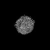
| Method | ELECTRON MICROSCOPY / single particle reconstruction / cryo EM / Resolution: 3.4 Å | ||||||||||||||||||||||||
 Authors Authors | Li, W. / Liu, B. / Cao, Q. | ||||||||||||||||||||||||
| Funding support |  United States, 7items United States, 7items
| ||||||||||||||||||||||||
 Citation Citation | Journal: Nature / Year: 2025 Title: Molecular basis of vitamin-K-driven γ-carboxylation at the membrane interface. Authors: Qing Cao / Aaron Ammerman / Mierxiati Saimi / Zongtao Lin / Guomin Shen / Huaping Chen / Jie Sun / Mengqi Chai / Shixuan Liu / Fong-Fu Hsu / Andrzej M Krezel / Michael L Gross / Jinbin Xu / ...Authors: Qing Cao / Aaron Ammerman / Mierxiati Saimi / Zongtao Lin / Guomin Shen / Huaping Chen / Jie Sun / Mengqi Chai / Shixuan Liu / Fong-Fu Hsu / Andrzej M Krezel / Michael L Gross / Jinbin Xu / Benjamin A Garcia / Bin Liu / Weikai Li /  Abstract: The γ-carboxylation of glutamate residues enables Ca-mediated membrane assembly of protein complexes that support broad physiological functions, including haemostasis, calcium homeostasis, immune ...The γ-carboxylation of glutamate residues enables Ca-mediated membrane assembly of protein complexes that support broad physiological functions, including haemostasis, calcium homeostasis, immune response and endocrine regulation. Modulating γ-carboxylation levels provides prevalent treatments for haemorrhagic and thromboembolic diseases. This unique post-translational modification requires vitamin K hydroquinone (KH) to drive highly demanding reactions catalysed by the membrane-integrated γ-carboxylase (VKGC). Here, to decipher the underlying mechanisms, we determined cryo-electron microscopy structures of human VKGC in unbound form, with KH and four haemostatic and non-haemostatic proteins possessing propeptides and glutamate-rich domains in different carboxylation states. VKGC recognizes substrate proteins through knob-and-hole interactions with propeptides, thereby bringing tethered glutamate-containing segments for processive carboxylation within a large chamber that provides steric control. Propeptide binding also triggers a global conformational change to signal VKGC activation. Through sequential deprotonation and KH epoxidation, VKGC generates a free hydroxide ion as an exceptionally strong base that is required to deprotonate the γ-carbon of glutamate for CO addition. The diffusion of this superbase-protected and guided by a sealed hydrophobic tunnel-elegantly resolves the challenge of coupling KH epoxidation to γ-carboxylation across the membrane interface. These structural insights and extensive functional experiments advance membrane enzymology and propel the development of treatments for γ-carboxylation disorders. | ||||||||||||||||||||||||
| History |
|
- Structure visualization
| Structure viewer | Molecule: MolmilJmol/JSmol |
|---|
- Downloads & links
-Download
| PDBx/mmCIF format | 9bvm.cif.gz | 171.8 KB | Display | PDBx/mmCIF format |
|---|---|---|---|---|
| PDB format | pdb9bvm.ent.gz | Display | PDB format | |
| PDBx/mmJSON format | 9bvm.json.gz | Tree view | PDBx/mmJSON format | |
| Others |  Other downloads Other downloads |
-Validation report
| Arichive directory | https://data.pdbj.org/pub/pdb/validation_reports/bv/9bvmftp://data.pdbj.org/pub/pdb/validation_reports/bv/9bvm | HTTPS FTP |
|---|
-Related structure data
| Related structure data |  44937MC  9bvkC  9bvlC  9bvoC 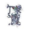 9bvpC 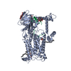 9bvqC  9bvrC M: map data used to model this data C: citing same article ( |
|---|---|
| Similar structure data |


-Links
 PDBj
PDBj


- Assembly
| Deposited unit | 
|
|---|---|
| 1 |
|
-Components
-Protein , 2 types, 2 molecules AP
| #1: Protein | Mass: 85005.680 Da / Num. of mol.: 1 Source method: isolated from a genetically manipulated source Source: (gene. exp.) Homo sapiens (human) / Gene: GGCX, GC / Cell line (production host): HEK293S / Production host: Homo sapiens (human)References: UniProt: P38435, peptidyl-glutamate 4-carboxylase |
|---|---|
| #2: Protein | Mass: 8219.110 Da / Num. of mol.: 1 Source method: isolated from a genetically manipulated source Source: (gene. exp.) Homo sapiens (human) / Gene: PROC / Cell line (production host): HEK293S / Production host: Homo sapiens (human) / References: UniProt: P04070 |
-Sugars , 2 types, 4 molecules 
| #3: Polysaccharide | Source method: isolated from a genetically manipulated source #6: Sugar | ChemComp-NAG / |  Type: D-saccharide, beta linking / Mass: 221.208 Da / Num. of mol.: 1 Type: D-saccharide, beta linking / Mass: 221.208 Da / Num. of mol.: 1Source method: isolated from a genetically manipulated source Formula: C8H15NO6 |
|---|
-Non-polymers , 2 types, 3 molecules 
| #4: Chemical | ChemComp-A1AVC / Mass: 452.712 Da / Num. of mol.: 1 / Source method: obtained synthetically / Formula: C31H48O2 / Feature type: SUBJECT OF INVESTIGATION |
|---|---|
| #5: Chemical |  Mass: 763.100 Da / Num. of mol.: 2 / Source method: obtained synthetically / Formula: C42H85NO8P / Feature type: SUBJECT OF INVESTIGATION / Comment: phospholipid*YM Mass: 763.100 Da / Num. of mol.: 2 / Source method: obtained synthetically / Formula: C42H85NO8P / Feature type: SUBJECT OF INVESTIGATION / Comment: phospholipid*YM |
-Details
| Has ligand of interest | Y |
|---|---|
| Has protein modification | Y |
-Experimental details
-Experiment
| Experiment | Method: ELECTRON MICROSCOPY |
|---|---|
| EM experiment | Aggregation state: PARTICLE / 3D reconstruction method: single particle reconstruction |
- Sample preparation
| Component | Name: Vitamin K-dependent gamma-carboxylase with protein C propeptide and glutamate-rich region and with vitamin K hydroquinone Type: COMPLEX / Entity ID: #1-#2 / Source: RECOMBINANT | ||||||||||||||||||||||||||||||
|---|---|---|---|---|---|---|---|---|---|---|---|---|---|---|---|---|---|---|---|---|---|---|---|---|---|---|---|---|---|---|---|
| Molecular weight | Value: 0.11 MDa / Experimental value: NO | ||||||||||||||||||||||||||||||
| Source (natural) | Organism: Homo sapiens (human) | ||||||||||||||||||||||||||||||
| Source (recombinant) | Organism: Homo sapiens (human) / Cell: HEK293S / Plasmid: pEG BacMam | ||||||||||||||||||||||||||||||
| Buffer solution | pH: 7.5 | ||||||||||||||||||||||||||||||
| Buffer component |
| ||||||||||||||||||||||||||||||
| Specimen | Embedding applied: NO / Shadowing applied: NO / Staining applied: NO / Vitrification applied: YES | ||||||||||||||||||||||||||||||
| Specimen support | Grid material: COPPER / Grid mesh size: 300 divisions/in. / Grid type: Quantifoil R1.2/1.3 | ||||||||||||||||||||||||||||||
| Vitrification | Instrument: LEICA EM GP / Cryogen name: ETHANE / Humidity: 95 % / Chamber temperature: 283.15 K |
- Electron microscopy imaging
| Experimental equipment |  Model: Titan Krios / Image courtesy: FEI Company |
|---|---|
| Microscopy | Model: TFS KRIOS |
| Electron gun | Electron source:  FIELD EMISSION GUN / Accelerating voltage: 300 kV / Illumination mode: FLOOD BEAM FIELD EMISSION GUN / Accelerating voltage: 300 kV / Illumination mode: FLOOD BEAM |
| Electron lens | Mode: BRIGHT FIELD / Nominal magnification: 130000 X / Nominal defocus max: 2000 nm / Nominal defocus min: 1000 nm / Cs: 2.7 mm / C2 aperture diameter: 50 µm / Alignment procedure: COMA FREE |
| Specimen holder | Cryogen: NITROGEN / Specimen holder model: FEI TITAN KRIOS AUTOGRID HOLDER / Temperature (max): 77 K / Temperature (min): 63 K |
| Image recording | Electron dose: 54.8 e/Å2 / Film or detector model: GATAN K3 BIOQUANTUM (6k x 4k) / Num. of grids imaged: 1 / Num. of real images: 5313 |
| EM imaging optics | Energyfilter name: GIF Bioquantum / Energyfilter slit width: 20 eV |
| Image scans | Width: 4092 / Height: 5760 |
- Processing
| EM software | Name: PHENIX / Version: 1.21.1_5286: / Category: model refinement | ||||||||||||||||||||||||
|---|---|---|---|---|---|---|---|---|---|---|---|---|---|---|---|---|---|---|---|---|---|---|---|---|---|
| CTF correction | Type: PHASE FLIPPING AND AMPLITUDE CORRECTION | ||||||||||||||||||||||||
| Particle selection | Num. of particles selected: 1322129 | ||||||||||||||||||||||||
| 3D reconstruction | Resolution: 3.4 Å / Resolution method: FSC 0.143 CUT-OFF / Num. of particles: 177459 / Symmetry type: POINT | ||||||||||||||||||||||||
| Atomic model building | Protocol: OTHER / Space: REAL | ||||||||||||||||||||||||
| Refine LS restraints |
|
