 Movie
Movie Controller

[English] 日本語
 Yorodumi
- PDB-9bth: Rhesus Fab 42056-a.01 in complex with CAP256SU.wk34 RnS SOSIP Env -
+ Open data

- Basic information
| Entry | Database: PDB / ID: 9bth | |||||||||||||||||||||||||||||||||
|---|---|---|---|---|---|---|---|---|---|---|---|---|---|---|---|---|---|---|---|---|---|---|---|---|---|---|---|---|---|---|---|---|---|---|
| Title | Rhesus Fab 42056-a.01 in complex with CAP256SU.wk34 RnS SOSIP Env | |||||||||||||||||||||||||||||||||
 Components Components |
| |||||||||||||||||||||||||||||||||
 Keywords Keywords | VIRAL PROTEIN/IMMUNE SYSTEM / HIV-1 / SOSIP / Vaccine / VIRAL PROTEIN-IMMUNE SYSTEM complex / V2-Apex / multi-donor / SHIV / IMMUNE SYSTEM | |||||||||||||||||||||||||||||||||
| Function / homology |  Function and homology information Function and homology informationsymbiont-mediated perturbation of host defense response / positive regulation of plasma membrane raft polarization / positive regulation of receptor clustering / host cell endosome membrane / clathrin-dependent endocytosis of virus by host cell / viral protein processing / fusion of virus membrane with host plasma membrane / fusion of virus membrane with host endosome membrane / viral envelope / virion attachment to host cell ...symbiont-mediated perturbation of host defense response / positive regulation of plasma membrane raft polarization / positive regulation of receptor clustering / host cell endosome membrane / clathrin-dependent endocytosis of virus by host cell / viral protein processing / fusion of virus membrane with host plasma membrane / fusion of virus membrane with host endosome membrane / viral envelope / virion attachment to host cell / host cell plasma membrane / virion membrane / structural molecule activity / membrane Similarity search - Function | |||||||||||||||||||||||||||||||||
| Biological species |    Human immunodeficiency virus 1 Human immunodeficiency virus 1 | |||||||||||||||||||||||||||||||||
| Method | ELECTRON MICROSCOPY / single particle reconstruction / cryo EM / Resolution: 4.2 Å | |||||||||||||||||||||||||||||||||
 Authors Authors | Gorman, J. / Kwong, P.D. | |||||||||||||||||||||||||||||||||
| Funding support |  United States, 2items United States, 2items
| |||||||||||||||||||||||||||||||||
 Citation Citation | Journal: J Exp Med / Year: 2025 Title: Structural and genetic basis of HIV-1 envelope V2 apex recognition by rhesus broadly neutralizing antibodies. Authors: Ryan S Roark / Rumi Habib / Jason Gorman / Hui Li / Andrew Jesse Connell / Mattia Bonsignori / Yicheng Guo / Michael P Hogarty / Adam S Olia / Kirsten J Sowers / Baoshan Zhang / Frederic ...Authors: Ryan S Roark / Rumi Habib / Jason Gorman / Hui Li / Andrew Jesse Connell / Mattia Bonsignori / Yicheng Guo / Michael P Hogarty / Adam S Olia / Kirsten J Sowers / Baoshan Zhang / Frederic Bibollet-Ruche / Tatsiana Bylund / Sean Callaghan / John W Carey / Gabriele Cerutti / Darcy R Harris / Wanting He / Emily Lewis / Tracy Liu / Rosemarie D Mason / Yujie Qiao / Younghoon Park / Juliette M Rando / Ajay Singh / Jeremy J Wolff / Q Paula Lei / Mark K Louder / Raiees Andrabi / Nicole A Doria-Rose / Kevin O Saunders / Michael S Seaman / Barton F Haynes / Daniel W Kulp / John R Mascola / Mario Roederer / Theodore C Pierson / Zizhang Sheng / Beatrice H Hahn / George M Shaw / Peter D Kwong / Lawrence Shapiro / Abstract: Broadly neutralizing antibodies targeting the V2 apex of HIV-1 envelope are desired as vaccine design templates, but few have been described. Here, we report 11 lineages of V2 apex-neutralizing ...Broadly neutralizing antibodies targeting the V2 apex of HIV-1 envelope are desired as vaccine design templates, but few have been described. Here, we report 11 lineages of V2 apex-neutralizing antibodies from simian-human immunodeficiency virus (SHIV)-infected rhesus macaques and determine cryo-EM structures for 9. A single V2 apex-neutralizing lineage accounted for cross-clade breadth in most macaques, and somatic hypermutation relative to breadth was generally low, exemplified by antibody V033-a.01 with <5% nucleotide mutation and 37% breadth (208-strain panel). Envelope complex structures revealed eight different antibody classes (one multi-donor) and the complete repertoire of all five possible recognition topologies, recapitulating canonical human modes of apex insertion and C-strand hydrogen bonding. Despite this diversity in recognition, all rhesus-V2 apex antibodies were derived from reading frame two of the DH3-15*01 gene. Collectively, these results define-in rhesus-the structural and genetic basis of HIV-1 V2 apex recognition and demonstrate unprecedented structural plasticity of a highly selected immunogenetic element. | |||||||||||||||||||||||||||||||||
| History |
|
- Structure visualization
| Structure viewer | Molecule: MolmilJmol/JSmol |
|---|
- Downloads & links
-Download
| PDBx/mmCIF format | 9bth.cif.gz | 393.5 KB | Display | PDBx/mmCIF format |
|---|---|---|---|---|
| PDB format | pdb9bth.ent.gz | 315.9 KB | Display | PDB format |
| PDBx/mmJSON format | 9bth.json.gz | Tree view | PDBx/mmJSON format | |
| Others |  Other downloads Other downloads |
-Validation report
| Arichive directory | https://data.pdbj.org/pub/pdb/validation_reports/bt/9bthftp://data.pdbj.org/pub/pdb/validation_reports/bt/9bth | HTTPS FTP |
|---|
-Related structure data
| Related structure data |  44890MC  9bnkC  9bnlC  9bnmC  9bnpC  9btiC  9btjC 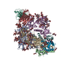 9btlC  9btvC M: map data used to model this data C: citing same article ( |
|---|---|
| Similar structure data |


-Links
 PDBj
PDBj


- Assembly
| Deposited unit | 
|
|---|---|
| 1 |
|
-Components
-Envelope glycoprotein ... , 2 types, 6 molecules GDFCEB
| #3: Protein | Mass: 53322.434 Da / Num. of mol.: 3 Source method: isolated from a genetically manipulated source Source: (gene. exp.) Human immunodeficiency virus 1 / Gene: env / Production host:  Homo sapiens (human) / References: UniProt: A0A0N9FF17 Homo sapiens (human) / References: UniProt: A0A0N9FF17#4: Protein | Mass: 17355.703 Da / Num. of mol.: 3 / Fragment: UNP residues 498-651 Source method: isolated from a genetically manipulated source Source: (gene. exp.) Human immunodeficiency virus 1 / Gene: env / Production host: Homo sapiens (human) / References: UniProt: A0A0N9FF17 |
|---|
-Antibody , 2 types, 2 molecules HL
| #1: Antibody | Mass: 26095.193 Da / Num. of mol.: 1 Source method: isolated from a genetically manipulated source Source: (gene. exp.) Human immunodeficiency virus 1 |
|---|---|
| #2: Antibody | Mass: 24365.133 Da / Num. of mol.: 1 Source method: isolated from a genetically manipulated source Source: (gene. exp.) Homo sapiens (human) |
-Sugars , 7 types, 70 molecules 
| #5: Polysaccharide | Source method: isolated from a genetically manipulated source #6: Polysaccharide | 2-acetamido-2-deoxy-beta-D-glucopyranose-(1-4)-2-acetamido-2-deoxy-beta-D-glucopyranose Source method: isolated from a genetically manipulated source #7: Polysaccharide | Source method: isolated from a genetically manipulated source #8: Polysaccharide | alpha-D-mannopyranose-(1-6)-alpha-D-mannopyranose-(1-6)-[alpha-D-mannopyranose-(1-3)]beta-D- ...alpha-D-mannopyranose-(1-6)-alpha-D-mannopyranose-(1-6)-[alpha-D-mannopyranose-(1-3)]beta-D-mannopyranose-(1-4)-2-acetamido-2-deoxy-beta-D-glucopyranose-(1-4)-2-acetamido-2-deoxy-beta-D-glucopyranose | Source method: isolated from a genetically manipulated source #9: Polysaccharide | alpha-D-mannopyranose-(1-2)-alpha-D-mannopyranose-(1-6)-[alpha-D-mannopyranose-(1-3)]alpha-D- ...alpha-D-mannopyranose-(1-2)-alpha-D-mannopyranose-(1-6)-[alpha-D-mannopyranose-(1-3)]alpha-D-mannopyranose-(1-6)-[alpha-D-mannopyranose-(1-2)-alpha-D-mannopyranose-(1-3)]beta-D-mannopyranose-(1-4)-2-acetamido-2-deoxy-beta-D-glucopyranose-(1-4)-2-acetamido-2-deoxy-beta-D-glucopyranose | Source method: isolated from a genetically manipulated source #10: Polysaccharide | alpha-D-mannopyranose-(1-2)-alpha-D-mannopyranose-(1-3)-[alpha-D-mannopyranose-(1-6)]alpha-D- ...alpha-D-mannopyranose-(1-2)-alpha-D-mannopyranose-(1-3)-[alpha-D-mannopyranose-(1-6)]alpha-D-mannopyranose-(1-6)-beta-D-mannopyranose-(1-4)-2-acetamido-2-deoxy-beta-D-glucopyranose-(1-4)-2-acetamido-2-deoxy-beta-D-glucopyranose | Type: oligosaccharide / Mass: 1235.105 Da / Num. of mol.: 1 Source method: isolated from a genetically manipulated source #11: Sugar | ChemComp-NAG /  Type: D-saccharide, beta linking / Mass: 221.208 Da / Num. of mol.: 56 / Source method: obtained synthetically / Formula: C8H15NO6 Type: D-saccharide, beta linking / Mass: 221.208 Da / Num. of mol.: 56 / Source method: obtained synthetically / Formula: C8H15NO6 |
|---|
-Details
| Has ligand of interest | N |
|---|---|
| Has protein modification | Y |
-Experimental details
-Experiment
| Experiment | Method: ELECTRON MICROSCOPY |
|---|---|
| EM experiment | Aggregation state: PARTICLE / 3D reconstruction method: single particle reconstruction |
- Sample preparation
| Component | Name: Rhesus Fab 42056-a.01 in complex with CAP256SU.wk34 RnS SOSIP Env Type: COMPLEX / Entity ID: #1-#4 / Source: RECOMBINANT |
|---|---|
| Molecular weight | Experimental value: NO |
| Source (natural) | Organism: Human immunodeficiency virus 1 |
| Source (recombinant) | Organism: Homo sapiens (human) |
| Buffer solution | pH: 7.4 / Details: PBS |
| Specimen | Conc.: 2 mg/ml / Embedding applied: NO / Shadowing applied: NO / Staining applied: NO / Vitrification applied: YES |
| Specimen support | Grid type: C-flat-1.2/1.3 |
| Vitrification | Instrument: FEI VITROBOT MARK IV / Cryogen name: ETHANE / Humidity: 90 % / Chamber temperature: 293 K |
- Electron microscopy imaging
| Experimental equipment |  Model: Titan Krios / Image courtesy: FEI Company |
|---|---|
| Microscopy | Model: FEI TITAN KRIOS |
| Electron gun | Electron source:  FIELD EMISSION GUN / Accelerating voltage: 300 kV / Illumination mode: FLOOD BEAM FIELD EMISSION GUN / Accelerating voltage: 300 kV / Illumination mode: FLOOD BEAM |
| Electron lens | Mode: BRIGHT FIELD / Nominal defocus max: 2500 nm / Nominal defocus min: 1000 nm / C2 aperture diameter: 70 µm |
| Image recording | Average exposure time: 2 sec. / Electron dose: 56.03 e/Å2 / Detector mode: COUNTING / Film or detector model: GATAN K3 (6k x 4k) / Num. of grids imaged: 1 |
- Processing
| EM software | Name: PHENIX / Version: dev_5278 / Category: model refinement |
|---|---|
| CTF correction | Type: PHASE FLIPPING AND AMPLITUDE CORRECTION |
| Symmetry | Point symmetry: C1 (asymmetric) |
| 3D reconstruction | Resolution: 4.2 Å / Resolution method: FSC 0.143 CUT-OFF / Num. of particles: 69264 / Symmetry type: POINT |
| Atomic model building | Protocol: FLEXIBLE FIT / Space: REAL |
| Atomic model building | PDB-ID: 6VTT Accession code: 6VTT / Source name: PDB / Type: experimental model |
| Refinement | Cross valid method: NONE |

