 Movie
Movie Controller


+ Open data

- Basic information
| Entry | Database: PDB / ID: 9bsh | ||||||
|---|---|---|---|---|---|---|---|
| Title | Staphylococcus aureus exfoliative toxin A D164A variant | ||||||
 Components Components | Exfoliative toxin A | ||||||
 Keywords Keywords | TOXIN / exfoliative toxin | ||||||
| Function / homology |  Function and homology information Function and homology informationsymbiont-mediated disruption of host cell-cell adhesion / Hydrolases; Acting on peptide bonds (peptidases); Serine endopeptidases / toxin activity / serine-type endopeptidase activity / proteolysis Similarity search - Function | ||||||
| Biological species |   Staphylococcus aureus (bacteria) Staphylococcus aureus (bacteria) | ||||||
| Method |  X-RAY DIFFRACTION / SYNCHROTRON / MOLECULAR REPLACEMENT / Resolution: 1.35 Å X-RAY DIFFRACTION / SYNCHROTRON / MOLECULAR REPLACEMENT / Resolution: 1.35 Å | ||||||
 Authors Authors | Holyoak, T. / Tran, N. | ||||||
| Funding support |  Canada, 1items Canada, 1items
| ||||||
 Citation Citation | Journal: Sci Adv / Year: 2025 Title: Identifying and controlling inactive and active conformations of a serine protease. Authors: Lee, E. / Tran, N. / Redzic, J.S. / Singh, H. / Alamillo, L. / Holyoak, T. / Hamelberg, D. / Eisenmesser, E.Z. | ||||||
| History |
|
- Structure visualization
| Structure viewer | Molecule: MolmilJmol/JSmol |
|---|
- Downloads & links
-Download
| PDBx/mmCIF format | 9bsh.cif.gz | 237.6 KB | Display | PDBx/mmCIF format |
|---|---|---|---|---|
| PDB format | pdb9bsh.ent.gz | 191.1 KB | Display | PDB format |
| PDBx/mmJSON format | 9bsh.json.gz | Tree view | PDBx/mmJSON format | |
| Others |  Other downloads Other downloads |
-Validation report
| Summary document | 9bsh_validation.pdf.gz | 435.1 KB | Display | wwPDB validaton report |
|---|---|---|---|---|
| Full document | 9bsh_full_validation.pdf.gz | 438 KB | Display | |
| Data in XML | 9bsh_validation.xml.gz | 34 KB | Display | |
| Data in CIF | 9bsh_validation.cif.gz | 50.6 KB | Display | |
| Arichive directory | https://data.pdbj.org/pub/pdb/validation_reports/bs/9bshftp://data.pdbj.org/pub/pdb/validation_reports/bs/9bsh | HTTPS FTP |
-Related structure data

-Links
 PDBj
PDBj- Assembly
| Deposited unit | 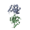
| ||||||||
|---|---|---|---|---|---|---|---|---|---|
| 1 | 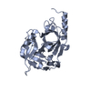
| ||||||||
| 2 | 
| ||||||||
| Unit cell |
|
-Components
| #1: Protein | Mass: 31091.061 Da / Num. of mol.: 2 / Mutation: D202A Source method: isolated from a genetically manipulated source Source: (gene. exp.) Staphylococcus aureus (bacteria) / Gene: eta / Production host: References: UniProt: P09331, Hydrolases; Acting on peptide bonds (peptidases); Serine endopeptidases #2: Water | ChemComp-HOH / |  Mass: 18.015 Da / Num. of mol.: 907 / Source method: isolated from a natural source / Formula: H2O Mass: 18.015 Da / Num. of mol.: 907 / Source method: isolated from a natural source / Formula: H2OHas protein modification | N | |
|---|
-Experimental details
-Experiment
| Experiment | Method: X-RAY DIFFRACTION / Number of used crystals: 1 |
|---|
- Sample preparation
| Crystal | Density Matthews: 2.12 Å3/Da / Density % sol: 41.79 % |
|---|---|
| Crystal grow | Temperature: 298 K / Method: vapor diffusion, hanging drop Details: 0.1M magnesium formate, 17.5% PEG 3350 (MCSG1 G10), 2:2 uL protein:mother liquor with a protein concentration of 17.5 mg/mL |
-Data collection
| Diffraction | Mean temperature: 100 K / Serial crystal experiment: N |
|---|---|
| Diffraction source | Source: SYNCHROTRON / Site: CLSI / Beamline: 08B1-1 / Wavelength: 1.18069 Å |
| Detector | Type: DECTRIS PILATUS3 6M / Detector: PIXEL / Date: Aug 16, 2023 |
| Radiation | Protocol: SINGLE WAVELENGTH / Monochromatic (M) / Laue (L): M / Scattering type: x-ray |
| Radiation wavelength | Wavelength: 1.18069 Å / Relative weight: 1 |
| Reflection | Resolution: 1.35→66.62 Å / Num. obs: 109787 / % possible obs: 96.38 % / Redundancy: 6.2 % / CC1/2: 0.996 / Rmerge(I) obs: 0.08 / Rpim(I) all: 0.037 / Rrim(I) all: 0.088 / Net I/σ(I): 21.8 |
| Reflection shell | Resolution: 1.35→1.37 Å / Redundancy: 5.8 % / Rmerge(I) obs: 0.344 / Mean I/σ(I) obs: 6.5 / Num. unique obs: 5330 / CC1/2: 0.675 / Rpim(I) all: 0.16 / Rrim(I) all: 0.381 / % possible all: 95.3 |
- Processing
| Software |
| |||||||||||||||||||||||||||||||||||||||||||||||||||||||||||||||||||||||||||||||||||||||||||||||||||||||||||||||||||||||||||||||||||||||||||||||||||||||||||||||||||||||||||||||||||||||||||||||||||||||||||||||||||||||||
|---|---|---|---|---|---|---|---|---|---|---|---|---|---|---|---|---|---|---|---|---|---|---|---|---|---|---|---|---|---|---|---|---|---|---|---|---|---|---|---|---|---|---|---|---|---|---|---|---|---|---|---|---|---|---|---|---|---|---|---|---|---|---|---|---|---|---|---|---|---|---|---|---|---|---|---|---|---|---|---|---|---|---|---|---|---|---|---|---|---|---|---|---|---|---|---|---|---|---|---|---|---|---|---|---|---|---|---|---|---|---|---|---|---|---|---|---|---|---|---|---|---|---|---|---|---|---|---|---|---|---|---|---|---|---|---|---|---|---|---|---|---|---|---|---|---|---|---|---|---|---|---|---|---|---|---|---|---|---|---|---|---|---|---|---|---|---|---|---|---|---|---|---|---|---|---|---|---|---|---|---|---|---|---|---|---|---|---|---|---|---|---|---|---|---|---|---|---|---|---|---|---|---|---|---|---|---|---|---|---|---|---|---|---|---|---|---|---|---|
| Refinement | Method to determine structure: MOLECULAR REPLACEMENT / Resolution: 1.35→51.57 Å / SU ML: 0.17 / Cross valid method: FREE R-VALUE / σ(F): 1.35 / Phase error: 22.37 / Stereochemistry target values: ML
| |||||||||||||||||||||||||||||||||||||||||||||||||||||||||||||||||||||||||||||||||||||||||||||||||||||||||||||||||||||||||||||||||||||||||||||||||||||||||||||||||||||||||||||||||||||||||||||||||||||||||||||||||||||||||
| Solvent computation | Shrinkage radii: 0.9 Å / VDW probe radii: 1.1 Å / Solvent model: FLAT BULK SOLVENT MODEL | |||||||||||||||||||||||||||||||||||||||||||||||||||||||||||||||||||||||||||||||||||||||||||||||||||||||||||||||||||||||||||||||||||||||||||||||||||||||||||||||||||||||||||||||||||||||||||||||||||||||||||||||||||||||||
| Refinement step | Cycle: LAST / Resolution: 1.35→51.57 Å
| |||||||||||||||||||||||||||||||||||||||||||||||||||||||||||||||||||||||||||||||||||||||||||||||||||||||||||||||||||||||||||||||||||||||||||||||||||||||||||||||||||||||||||||||||||||||||||||||||||||||||||||||||||||||||
| Refine LS restraints |
| |||||||||||||||||||||||||||||||||||||||||||||||||||||||||||||||||||||||||||||||||||||||||||||||||||||||||||||||||||||||||||||||||||||||||||||||||||||||||||||||||||||||||||||||||||||||||||||||||||||||||||||||||||||||||
| LS refinement shell |
| |||||||||||||||||||||||||||||||||||||||||||||||||||||||||||||||||||||||||||||||||||||||||||||||||||||||||||||||||||||||||||||||||||||||||||||||||||||||||||||||||||||||||||||||||||||||||||||||||||||||||||||||||||||||||
| Refinement TLS params. | Method: refined / Origin x: 12.9634 Å / Origin y: -17.201 Å / Origin z: 20.5404 Å
| |||||||||||||||||||||||||||||||||||||||||||||||||||||||||||||||||||||||||||||||||||||||||||||||||||||||||||||||||||||||||||||||||||||||||||||||||||||||||||||||||||||||||||||||||||||||||||||||||||||||||||||||||||||||||
| Refinement TLS group | Selection details: all |
