[English] 日本語
Yorodumi
- PDB-9brh: Crystal Structure of Human G Protein-Coupled Receptor Kinase 5 in... -

+
Open data


ID or keywords:

Loading...

-
Basic information

Entry
Database: PDB / ID: 9brh
Title
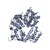
Crystal Structure of Human G Protein-Coupled Receptor Kinase 5 in Complex with GRL056-21
ComponentsG protein-coupled receptor kinase 5
KeywordsSIGNALING PROTEIN/INHIBITOR / G protein-coupled receptor / GPCR G protein-coupled receptor kinase / GRK kinase / SIGNALING PROTEIN-INHIBITOR complex
Function / homology
Function and homology information


G-protein-coupled receptor kinase / beta-adrenergic receptor kinase activity / G protein-coupled receptor kinase activity / tachykinin receptor signaling pathway / regulation of G protein-coupled receptor signaling pathway / fat cell differentiation / regulation of signal transduction / protein kinase C binding / phospholipid binding / Wnt signaling pathway ...G-protein-coupled receptor kinase / beta-adrenergic receptor kinase activity / G protein-coupled receptor kinase activity / tachykinin receptor signaling pathway / regulation of G protein-coupled receptor signaling pathway / fat cell differentiation / regulation of signal transduction / protein kinase C binding / phospholipid binding / Wnt signaling pathway / adenylate cyclase-modulating G protein-coupled receptor signaling pathway / nuclear membrane / protein autophosphorylation / G alpha (s) signalling events / G alpha (q) signalling events / regulation of cell cycle / protein kinase activity / nuclear speck / G protein-coupled receptor signaling pathway / protein serine/threonine kinase activity / apoptotic process / positive regulation of cell population proliferation / negative regulation of apoptotic process / ATP binding / plasma membrane / cytosol / cytoplasm
Similarity search - Function
GPCR kinase / Regulator of G protein signaling domain / RGS domain / RGS domain profile. / Regulator of G protein signalling domain / RGS, subdomain 2 / RGS domain superfamily / Extension to Ser/Thr-type protein kinases / AGC-kinase, C-terminal / AGC-kinase C-terminal domain profile. ...GPCR kinase / Regulator of G protein signaling domain / RGS domain / RGS domain profile. / Regulator of G protein signalling domain / RGS, subdomain 2 / RGS domain superfamily / Extension to Ser/Thr-type protein kinases / AGC-kinase, C-terminal / AGC-kinase C-terminal domain profile. / Protein kinase domain / Serine/Threonine protein kinases, catalytic domain / Protein kinase, ATP binding site / Protein kinases ATP-binding region signature. / Protein kinase domain profile. / Protein kinase domain / Protein kinase-like domain superfamily
Similarity search - Domain/homology
: / G protein-coupled receptor kinase 5
Similarity search - Component
Biological speciesHomo sapiens (human)
MethodX-RAY DIFFRACTION / SYNCHROTRON / MOLECULAR REPLACEMENT / Resolution: 3.69 Å
AuthorsChen, Y. / Tesmer, J.J.G.
Funding support United States, 2items
OrganizationGrant numberCountry
National Institutes of Health/National Heart, Lung, and Blood Institute (NIH/NHLBI)HL071818 United States
National Institutes of Health/National Center for Research Resources (NIH/NCRR)CA023168 United States
CitationJournal: Eur.J.Med.Chem. / Year: 2025
Title: Design, synthesis, and X-ray structural studies of a series of highly potent, selective, and drug-like G protein-coupled receptor kinase 5 inhibitors.
Authors: Ghosh, A.K. / Chen, Y. / Gadi, R.K. / Sonawane, A. / Gamage, S.P. / Tesmer, J.G.
History
DepositionMay 11, 2024Deposition site: RCSB / Processing site: RCSB
Revision 1.0Feb 19, 2025Provider: repository / Type: Initial release

-
Structure visualization

Structure viewerMolecule:
MolmilJmol/JSmol

Downloads & links

-
Assembly

Deposited unit
A: G protein-coupled receptor kinase 5
hetero molecules


Theoretical massNumber of molelcules
Total (without water)69,4442
Polymers68,9321
Non-polymers5131
Water181
1


  • Idetical with deposited unit
  • defined by author&software
TypeNameSymmetry operationNumber
identity operation1_555x,y,z1
Buried area0 Å2
ΔGint0 kcal/mol
Surface area25080 Å2
MethodPISA
Unit cell
Length a, b, c (Å)138.639, 138.639, 71.707
Angle α, β, γ (deg.)90.000, 90.000, 90.000
Int Tables number92
Space group name H-MP41212
Space group name HallP4abw2nw
Symmetry operation#1: x,y,z
#2: -y+1/2,x+1/2,z+1/4
#3: y+1/2,-x+1/2,z+3/4
#4: x+1/2,-y+1/2,-z+3/4
#5: -x+1/2,y+1/2,-z+1/4
#6: -x,-y,z+1/2
#7: y,x,-z
#8: -y,-x,-z+1/2

-
Components

#1: Protein G protein-coupled receptor kinase 5


Mass: 68931.727 Da / Num. of mol.: 1 / Mutation: D311N
Source method: isolated from a genetically manipulated source
Source: (gene. exp.) Homo sapiens (human) / Gene: GRK5 / Production host: Escherichia coli BL21(DE3) (bacteria) / Strain (production host): Rosetta / References: UniProt: P34947
#2: Chemical ChemComp-A1AQ7 / (3Z)-N-[(1R)-1-(4-fluorophenyl)ethyl]-3-({4-[(furan-3-carbonyl)amino]-3,5-dimethyl-1H-pyrrol-2-yl}methylidene)-2-oxo-3,7-dihydro-2H-indole-5-carboxamide


Mass: 512.532 Da / Num. of mol.: 1 / Source method: obtained synthetically / Formula: C29H25FN4O4 / Feature type: SUBJECT OF INVESTIGATION
#3: Water ChemComp-HOH / water


Mass: 18.015 Da / Num. of mol.: 1 / Source method: isolated from a natural source / Formula: H2O
Has ligand of interestY
Has protein modificationN

-
Experimental details

-
Experiment

ExperimentMethod: X-RAY DIFFRACTION / Number of used crystals: 1

-
Sample preparation

CrystalDensity Matthews: 2.5 Å3/Da / Density % sol: 50.79 %
Crystal growTemperature: 277.15 K / Method: vapor diffusion, hanging drop
Details: 220 mM potassium citrate tribasic, 20% PEG3350, crystals soaked in 25% PEG3350, 10% glycerol, 1 mM inhibitor

-
Data collection

DiffractionMean temperature: 100 K / Serial crystal experiment: N
Diffraction sourceSource: SYNCHROTRON / Site: NSLS-II / Beamline: 17-ID-2 / Wavelength: 0.9201 Å
DetectorType: DECTRIS EIGER X 16M / Detector: PIXEL / Date: Jul 17, 2023
RadiationProtocol: SINGLE WAVELENGTH / Monochromatic (M) / Laue (L): M / Scattering type: x-ray
Radiation wavelengthWavelength: 0.9201 Å / Relative weight: 1
ReflectionResolution: 3.69→29.74 Å / Num. obs: 7927 / % possible obs: 99.7 % / Redundancy: 12.9 % / Biso Wilson estimate: 103.91 Å2 / CC1/2: 0.996 / Net I/σ(I): 9.4
Reflection shellResolution: 3.69→3.79 Å / Num. unique obs: 7162 / CC1/2: 0.802

-
Processing

Software
NameVersionClassification
PHENIX1.21.1_5286refinement
autoPROCdata reduction
Aimlessdata scaling
MOLREPphasing
RefinementMethod to determine structure: MOLECULAR REPLACEMENT / Resolution: 3.69→29.74 Å / SU ML: 0.4071 / Cross valid method: FREE R-VALUE / σ(F): 1.37 / Phase error: 23.7227
Stereochemistry target values: GeoStd + Monomer Library + CDL v1.2
RfactorNum. reflection% reflection
Rfree0.2445 790 10.01 %
Rwork0.1943 7102 -
obs0.1995 7892 99.86 %
Solvent computationShrinkage radii: 0.9 Å / VDW probe radii: 1.1 Å / Solvent model: FLAT BULK SOLVENT MODEL
Displacement parametersBiso mean: 119.64 Å2
Refinement stepCycle: LAST / Resolution: 3.69→29.74 Å
ProteinNucleic acidLigandSolventTotal
Num. atoms4092 0 38 1 4131
Refine LS restraints
Refine-IDTypeDev idealNumber
X-RAY DIFFRACTIONf_bond_d0.00264224
X-RAY DIFFRACTIONf_angle_d0.49235687
X-RAY DIFFRACTIONf_chiral_restr0.0376588
X-RAY DIFFRACTIONf_plane_restr0.0046742
X-RAY DIFFRACTIONf_dihedral_angle_d13.40191621
LS refinement shell
Resolution (Å)Rfactor RfreeNum. reflection RfreeRfactor RworkNum. reflection RworkRefine-ID% reflection obs (%)
3.69-3.920.271280.23891157X-RAY DIFFRACTION99.3
3.92-4.230.27821290.20871153X-RAY DIFFRACTION100
4.23-4.650.24231280.17691157X-RAY DIFFRACTION100
4.65-5.320.20531310.16971178X-RAY DIFFRACTION100
5.32-6.690.30751330.22761189X-RAY DIFFRACTION100
6.7-29.740.21761410.18051268X-RAY DIFFRACTION99.86
Refinement TLS params.Method: refined / Origin x: 32.114943588 Å / Origin y: 5.94426536597 Å / Origin z: 6.48755288804 Å
111213212223313233
T0.777700267056 Å20.100802930307 Å20.00340839375818 Å2-0.722084085216 Å20.112173826749 Å2--0.604975940066 Å2
L1.84175569168 °21.0146805456 °20.321269148613 °2-3.65300840928 °20.107388614381 °2--2.33251684951 °2
S-0.141156559831 Å °0.123120268852 Å °-0.0279630111626 Å °-0.421900634779 Å °0.102258331854 Å °-0.116642307949 Å °-0.258387851987 Å °0.0740136997351 Å °0.0367563651835 Å °
Refinement TLS groupSelection details: all

+
About Yorodumi

-
News

-
Feb 9, 2022. New format data for meta-information of EMDB entries

New format data for meta-information of EMDB entries

  • Version 3 of the EMDB header file is now the official format.
  • The previous official version 1.9 will be removed from the archive.

Related info.:EMDB header

External links:wwPDB to switch to version 3 of the EMDB data model

-
Aug 12, 2020. Covid-19 info

Covid-19 info

URL: https://pdbj.org/emnavi/covid19.php

New page: Covid-19 featured information page in EM Navigator.

Related info.:Covid-19 info / Mar 5, 2020. Novel coronavirus structure data

+
Mar 5, 2020. Novel coronavirus structure data

Novel coronavirus structure data

Related info.:Yorodumi Speices / Aug 12, 2020. Covid-19 info

External links:COVID-19 featured content - PDBj / Molecule of the Month (242):Coronavirus Proteases

+
Jan 31, 2019. EMDB accession codes are about to change! (news from PDBe EMDB page)

EMDB accession codes are about to change! (news from PDBe EMDB page)

  • The allocation of 4 digits for EMDB accession codes will soon come to an end. Whilst these codes will remain in use, new EMDB accession codes will include an additional digit and will expand incrementally as the available range of codes is exhausted. The current 4-digit format prefixed with “EMD-” (i.e. EMD-XXXX) will advance to a 5-digit format (i.e. EMD-XXXXX), and so on. It is currently estimated that the 4-digit codes will be depleted around Spring 2019, at which point the 5-digit format will come into force.
  • The EM Navigator/Yorodumi systems omit the EMD- prefix.

Related info.:Q: What is EMD? / ID/Accession-code notation in Yorodumi/EM Navigator

External links:EMDB Accession Codes are Changing Soon! / Contact to PDBj

+
Jul 12, 2017. Major update of PDB

Major update of PDB

  • wwPDB released updated PDB data conforming to the new PDBx/mmCIF dictionary.
  • This is a major update changing the version number from 4 to 5, and with Remediation, in which all the entries are updated.
  • In this update, many items about electron microscopy experimental information are reorganized (e.g. em_software).
  • Now, EM Navigator and Yorodumi are based on the updated data.

External links:wwPDB Remediation / Enriched Model Files Conforming to OneDep Data Standards Now Available in the PDB FTP Archive

-
Yorodumi

Thousand views of thousand structures

  • Yorodumi is a browser for structure data from EMDB, PDB, SASBDB, etc.
  • This page is also the successor to EM Navigator detail page, and also detail information page/front-end page for Omokage search.
  • The word "yorodu" (or yorozu) is an old Japanese word meaning "ten thousand". "mi" (miru) is to see.

Related info.:EMDB / PDB / SASBDB / Comparison of 3 databanks / Yorodumi Search / Aug 31, 2016. New EM Navigator & Yorodumi / Yorodumi Papers / Jmol/JSmol / Function and homology information / Changes in new EM Navigator and Yorodumi

Read more