 Movie
Movie Controller

[English] 日本語
 Yorodumi
- PDB-9blo: T450C mutant of repeat domain 2 from Clostridium perfringens adhe... -
+ Open data

- Basic information
| Entry | Database: PDB / ID: 9blo | ||||||
|---|---|---|---|---|---|---|---|
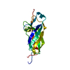
| Title | T450C mutant of repeat domain 2 from Clostridium perfringens adhesin CPE0147 without intramolecular ester bond | ||||||
 Components Components | Surface anchored protein | ||||||
 Keywords Keywords | UNKNOWN FUNCTION / repeat domain / adhesin / bacterial adhesion / Ig-like domain | ||||||
| Function / homology |  Function and homology information Function and homology information | ||||||
| Biological species |  Clostridium perfringens B str. ATCC 3626 (bacteria) Clostridium perfringens B str. ATCC 3626 (bacteria) | ||||||
| Method |  X-RAY DIFFRACTION / SYNCHROTRON / MOLECULAR REPLACEMENT / Resolution: 1.35 Å X-RAY DIFFRACTION / SYNCHROTRON / MOLECULAR REPLACEMENT / Resolution: 1.35 Å | ||||||
 Authors Authors | Squire, C.J. / Yosaatmadja, Y. | ||||||
| Funding support |  New Zealand, 1items New Zealand, 1items
| ||||||
 Citation Citation | Journal: Protein Sci. / Year: 2025 Title: Protease mimicry: Dissecting the ester bond crosslinking mechanics in bacterial adhesin proteins. Authors: Yosaatmadja, Y. / Ung, V. / Liu, X. / Zhao, Y. / Wardega, J.K. / Shetty, A. / Schoensee, S. / Leung, I.K.H. / Keown, J.R. / Goldstone, D.C. / Baker, E.N. / Young, P.G. / Mercadante, D. / Squire, C.J. | ||||||
| History |
|
- Structure visualization
| Structure viewer | Molecule: MolmilJmol/JSmol |
|---|
- Downloads & links
-Download
| PDBx/mmCIF format | 9blo.cif.gz | 132.9 KB | Display | PDBx/mmCIF format |
|---|---|---|---|---|
| PDB format | pdb9blo.ent.gz | 99 KB | Display | PDB format |
| PDBx/mmJSON format | 9blo.json.gz | Tree view | PDBx/mmJSON format | |
| Others |  Other downloads Other downloads |
-Validation report
| Summary document | 9blo_validation.pdf.gz | 427.4 KB | Display | wwPDB validaton report |
|---|---|---|---|---|
| Full document | 9blo_full_validation.pdf.gz | 427.2 KB | Display | |
| Data in XML | 9blo_validation.xml.gz | 17.7 KB | Display | |
| Data in CIF | 9blo_validation.cif.gz | 24.6 KB | Display | |
| Arichive directory | https://data.pdbj.org/pub/pdb/validation_reports/bl/9bloftp://data.pdbj.org/pub/pdb/validation_reports/bl/9blo | HTTPS FTP |
-Related structure data


-Links
 PDBj
PDBj- Assembly
| Deposited unit | 
| ||||||||
|---|---|---|---|---|---|---|---|---|---|
| 1 | 
| ||||||||
| 2 | 
| ||||||||
| Unit cell |
|
-Components
| #1: Protein | Mass: 17002.623 Da / Num. of mol.: 2 / Fragment: Repeat domain 2, residues 439-587 / Mutation: T450C Source method: isolated from a genetically manipulated source Source: (gene. exp.) Clostridium perfringens B str. ATCC 3626 (bacteria)Gene: AC1_0147 / Plasmid: pPROEXHTa / Production host: #2: Chemical |   Mass: 24.305 Da / Num. of mol.: 2 / Source method: obtained synthetically / Formula: Mg Mass: 24.305 Da / Num. of mol.: 2 / Source method: obtained synthetically / Formula: Mg#3: Chemical |   Mass: 40.078 Da / Num. of mol.: 2 / Source method: obtained synthetically / Formula: Ca Mass: 40.078 Da / Num. of mol.: 2 / Source method: obtained synthetically / Formula: Ca#4: Water | ChemComp-HOH / |  Mass: 18.015 Da / Num. of mol.: 283 / Source method: isolated from a natural source / Formula: H2O Mass: 18.015 Da / Num. of mol.: 283 / Source method: isolated from a natural source / Formula: H2OHas ligand of interest | N | Has protein modification | N | |
|---|
-Experimental details
-Experiment
| Experiment | Method: X-RAY DIFFRACTION / Number of used crystals: 1 |
|---|
- Sample preparation
| Crystal | Density Matthews: 2.26 Å3/Da / Density % sol: 45.46 % |
|---|---|
| Crystal grow | Temperature: 291 K / Method: vapor diffusion, sitting drop / pH: 8.5 Details: 0.2 M MgCl2, 0.1 M Tris.HCl pH 8.5, and 30% (w/v) PEG 4000 |
-Data collection
| Diffraction | Mean temperature: 100 K / Serial crystal experiment: N |
|---|---|
| Diffraction source | Source: SYNCHROTRON / Site: Australian Synchrotron  / Beamline: MX1 / Wavelength: 0.9537 Å / Beamline: MX1 / Wavelength: 0.9537 Å |
| Detector | Type: DECTRIS EIGER X 9M / Detector: PIXEL / Date: Oct 5, 2015 |
| Radiation | Protocol: SINGLE WAVELENGTH / Monochromatic (M) / Laue (L): M / Scattering type: x-ray |
| Radiation wavelength | Wavelength: 0.9537 Å / Relative weight: 1 |
| Reflection | Resolution: 1.35→18.85 Å / Num. obs: 61888 / % possible obs: 94.8 % / Redundancy: 6.7 % / CC1/2: 1 / Rpim(I) all: 0.023 / Net I/σ(I): 27.8 |
| Reflection shell | Resolution: 1.35→1.37 Å / Redundancy: 6.6 % / Mean I/σ(I) obs: 2.4 / Num. unique obs: 2494 / CC1/2: 0.787 / Rpim(I) all: 0.352 / % possible all: 77.4 |
- Processing
| Software |
| |||||||||||||||||||||||||||||||||||||||||||||||||||||||||||||||||||||||||||||||||||||||||||||||||||||||||||||||||||||||||||||||||||||||||||||||||||||||||||
|---|---|---|---|---|---|---|---|---|---|---|---|---|---|---|---|---|---|---|---|---|---|---|---|---|---|---|---|---|---|---|---|---|---|---|---|---|---|---|---|---|---|---|---|---|---|---|---|---|---|---|---|---|---|---|---|---|---|---|---|---|---|---|---|---|---|---|---|---|---|---|---|---|---|---|---|---|---|---|---|---|---|---|---|---|---|---|---|---|---|---|---|---|---|---|---|---|---|---|---|---|---|---|---|---|---|---|---|---|---|---|---|---|---|---|---|---|---|---|---|---|---|---|---|---|---|---|---|---|---|---|---|---|---|---|---|---|---|---|---|---|---|---|---|---|---|---|---|---|---|---|---|---|---|---|---|---|
| Refinement | Method to determine structure: MOLECULAR REPLACEMENT / Resolution: 1.35→18.85 Å / Cor.coef. Fo:Fc: 0.96 / Cor.coef. Fo:Fc free: 0.943 / SU B: 2.839 / SU ML: 0.051 / Cross valid method: FREE R-VALUE / ESU R: 0.074 / ESU R Free: 0.068 Details: Hydrogens have been added in their riding positions
| |||||||||||||||||||||||||||||||||||||||||||||||||||||||||||||||||||||||||||||||||||||||||||||||||||||||||||||||||||||||||||||||||||||||||||||||||||||||||||
| Solvent computation | Ion probe radii: 0.8 Å / Shrinkage radii: 0.8 Å / VDW probe radii: 1.2 Å / Solvent model: MASK BULK SOLVENT | |||||||||||||||||||||||||||||||||||||||||||||||||||||||||||||||||||||||||||||||||||||||||||||||||||||||||||||||||||||||||||||||||||||||||||||||||||||||||||
| Displacement parameters | Biso mean: 15.084 Å2
| |||||||||||||||||||||||||||||||||||||||||||||||||||||||||||||||||||||||||||||||||||||||||||||||||||||||||||||||||||||||||||||||||||||||||||||||||||||||||||
| Refinement step | Cycle: LAST / Resolution: 1.35→18.85 Å
| |||||||||||||||||||||||||||||||||||||||||||||||||||||||||||||||||||||||||||||||||||||||||||||||||||||||||||||||||||||||||||||||||||||||||||||||||||||||||||
| Refine LS restraints |
| |||||||||||||||||||||||||||||||||||||||||||||||||||||||||||||||||||||||||||||||||||||||||||||||||||||||||||||||||||||||||||||||||||||||||||||||||||||||||||
| LS refinement shell |
|
