 Movie
Movie Controller

[English] 日本語
 Yorodumi
- PDB-9bk8: Crystal structure of RidA family protein PA5083 from Pseudomonas ... -
+ Open data

- Basic information
| Entry | Database: PDB / ID: 9bk8 | ||||||
|---|---|---|---|---|---|---|---|
| Title | Crystal structure of RidA family protein PA5083 from Pseudomonas aeruginosa with 2-ketobutyric acid bound | ||||||
 Components Components | RidA family protein PA5083 | ||||||
 Keywords Keywords | HYDROLASE / RidA protein / deamination / stress | ||||||
| Function / homology | RutC family, YoaB-like / YjgF/YER057c/UK114 family / Endoribonuclease L-PSP / RutC-like superfamily / 2-KETOBUTYRIC ACID / RidA family protein Function and homology information Function and homology information | ||||||
| Biological species |   Pseudomonas aeruginosa (bacteria) Pseudomonas aeruginosa (bacteria) | ||||||
| Method |  X-RAY DIFFRACTION / MOLECULAR REPLACEMENT / Resolution: 1.67 Å X-RAY DIFFRACTION / MOLECULAR REPLACEMENT / Resolution: 1.67 Å | ||||||
 Authors Authors | Zhou, D. / Chen, L. / Rose, J.P. / Wang, B.C. | ||||||
| Funding support | 1items
| ||||||
 Citation Citation | Journal: To Be Published Title: Crystal structure of RidA family protein PA5083 from Pseudomonas aeruginosa with 2-ketobutyric acid bound Authors: Zhou, D. / Chen, L. / Rose, J.P. / Wang, B.C. | ||||||
| History |
|
- Structure visualization
| Structure viewer | Molecule: MolmilJmol/JSmol |
|---|
- Downloads & links
-Download
| PDBx/mmCIF format | 9bk8.cif.gz | 69.8 KB | Display | PDBx/mmCIF format |
|---|---|---|---|---|
| PDB format | pdb9bk8.ent.gz | 42.4 KB | Display | PDB format |
| PDBx/mmJSON format | 9bk8.json.gz | Tree view | PDBx/mmJSON format | |
| Others |  Other downloads Other downloads |
-Validation report
| Arichive directory | https://data.pdbj.org/pub/pdb/validation_reports/bk/9bk8ftp://data.pdbj.org/pub/pdb/validation_reports/bk/9bk8 | HTTPS FTP |
|---|
-Related structure data
| Related structure data | |
|---|---|
| Similar structure data |


-Links
 PDBj
PDBj
- Assembly
| Deposited unit | 
| |||||||||||||||||||||||||||
|---|---|---|---|---|---|---|---|---|---|---|---|---|---|---|---|---|---|---|---|---|---|---|---|---|---|---|---|---|
| 1 | 
| |||||||||||||||||||||||||||
| Unit cell |
| |||||||||||||||||||||||||||
| Components on special symmetry positions |
|
-Components
| #1: Protein | Mass: 12571.302 Da / Num. of mol.: 1 Source method: isolated from a genetically manipulated source Source: (gene. exp.) Pseudomonas aeruginosa (bacteria)Gene: yabJ_3, CAZ10_01035, GNQ48_03865, GUL26_07970, IPC1295_08400, IPC737_05390, NCTC13621_05579, PA52Ts2_0667 Production host: References: UniProt: A0A069Q0R8, Hydrolases; Acting on carbon-nitrogen bonds, other than peptide bonds; In cyclic amidines |
|---|---|
| #2: Chemical | ChemComp-2KT / 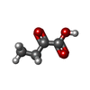  Mass: 102.089 Da / Num. of mol.: 1 / Source method: obtained synthetically / Formula: C4H6O3 / Feature type: SUBJECT OF INVESTIGATION Mass: 102.089 Da / Num. of mol.: 1 / Source method: obtained synthetically / Formula: C4H6O3 / Feature type: SUBJECT OF INVESTIGATION |
| #3: Water | ChemComp-HOH /  Mass: 18.015 Da / Num. of mol.: 120 / Source method: isolated from a natural source / Formula: H2O Mass: 18.015 Da / Num. of mol.: 120 / Source method: isolated from a natural source / Formula: H2O |
| Has ligand of interest | Y |
-Experimental details
-Experiment
| Experiment | Method: X-RAY DIFFRACTION / Number of used crystals: 1 |
|---|
- Sample preparation
| Crystal | Density Matthews: 3.05 Å3/Da / Density % sol: 59.71 % |
|---|---|
| Crystal grow | Temperature: 298 K / Method: vapor diffusion, hanging drop / Details: 0.1 M BIS-TRIS pH 5.5, 2.0 M Ammonium sulfate |
-Data collection
| Diffraction | Mean temperature: 100 K / Serial crystal experiment: N |
|---|---|
| Diffraction source | Source: ROTATING ANODE / Type: RIGAKU MICROMAX-002 / Wavelength: 1.5418 Å |
| Detector | Type: DECTRIS PILATUS 200K / Detector: PIXEL / Date: Jun 27, 2017 |
| Radiation | Protocol: SINGLE WAVELENGTH / Monochromatic (M) / Laue (L): M / Scattering type: x-ray |
| Radiation wavelength | Wavelength: 1.5418 Å / Relative weight: 1 |
| Reflection | Resolution: 1.67→50 Å / Num. obs: 17998 / % possible obs: 99.4 % / Redundancy: 6.2 % / Biso Wilson estimate: 17.86 Å2 / Rmerge(I) obs: 0.054 / Net I/σ(I): 36.43 |
| Reflection shell | Resolution: 1.67→1.7 Å / Rmerge(I) obs: 0.424 / Num. unique obs: 789 / CC1/2: 0.872 / % possible all: 89.1 |
- Processing
| Software |
| ||||||||||||||||||||||||||||||||||||||||||||||||||||||||||||||||||||||||||||||||||||||||||||||||||
|---|---|---|---|---|---|---|---|---|---|---|---|---|---|---|---|---|---|---|---|---|---|---|---|---|---|---|---|---|---|---|---|---|---|---|---|---|---|---|---|---|---|---|---|---|---|---|---|---|---|---|---|---|---|---|---|---|---|---|---|---|---|---|---|---|---|---|---|---|---|---|---|---|---|---|---|---|---|---|---|---|---|---|---|---|---|---|---|---|---|---|---|---|---|---|---|---|---|---|---|
| Refinement | Method to determine structure: MOLECULAR REPLACEMENT / Resolution: 1.67→37.77 Å / SU ML: 0.1675 / Cross valid method: FREE R-VALUE / σ(F): 1.34 / Phase error: 20.2934 Stereochemistry target values: GeoStd + Monomer Library + CDL v1.2
| ||||||||||||||||||||||||||||||||||||||||||||||||||||||||||||||||||||||||||||||||||||||||||||||||||
| Solvent computation | Shrinkage radii: 0.9 Å / VDW probe radii: 1.1 Å / Solvent model: FLAT BULK SOLVENT MODEL | ||||||||||||||||||||||||||||||||||||||||||||||||||||||||||||||||||||||||||||||||||||||||||||||||||
| Displacement parameters | Biso mean: 21.74 Å2 | ||||||||||||||||||||||||||||||||||||||||||||||||||||||||||||||||||||||||||||||||||||||||||||||||||
| Refinement step | Cycle: LAST / Resolution: 1.67→37.77 Å
| ||||||||||||||||||||||||||||||||||||||||||||||||||||||||||||||||||||||||||||||||||||||||||||||||||
| Refine LS restraints |
| ||||||||||||||||||||||||||||||||||||||||||||||||||||||||||||||||||||||||||||||||||||||||||||||||||
| LS refinement shell |
|
