| Entry | Database: PDB / ID: 9bja
|
|---|
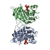
| Title | C. difficile Tcdb cysteine protease domain in complex with IP6 |
|---|
 Components Components | Toxin B |
|---|
 Keywords Keywords | TOXIN / catalytic activity peptidase activity catalytic activity / acting on a protein toxin activity small molecule binding |
|---|
| Function / homology |  Function and homology information Function and homology information
symbiont-mediated perturbation of host actin cytoskeleton via filamentous actin depolymerization / glucosyltransferase activity / Transferases; Glycosyltransferases; Hexosyltransferases / host cell cytosol / cysteine-type peptidase activity / host cell endosome membrane / toxin activity / Hydrolases; Acting on peptide bonds (peptidases); Cysteine endopeptidases / lipid binding / host cell plasma membrane ...symbiont-mediated perturbation of host actin cytoskeleton via filamentous actin depolymerization / glucosyltransferase activity / Transferases; Glycosyltransferases; Hexosyltransferases / host cell cytosol / cysteine-type peptidase activity / host cell endosome membrane / toxin activity / Hydrolases; Acting on peptide bonds (peptidases); Cysteine endopeptidases / lipid binding / host cell plasma membrane / proteolysis / extracellular region / metal ion bindingSimilarity search - Function TcdA/TcdB toxin, pore forming domain / TcdA/TcdB pore forming domain / CGT/MARTX, cysteine protease (CPD) domain / CGT/MARTX, cysteine protease (CPD) domain superfamily / Peptidase C80 family / CGT/MARTX cysteine protease (CPD) domain profile. / TcdA/TcdB toxin, N-terminal helical domain / TcdB toxin N-terminal helical domain / TcdA/TcdB toxin, catalytic glycosyltransferase domain / TcdA/TcdB catalytic glycosyltransferase domain ...TcdA/TcdB toxin, pore forming domain / TcdA/TcdB pore forming domain / CGT/MARTX, cysteine protease (CPD) domain / CGT/MARTX, cysteine protease (CPD) domain superfamily / Peptidase C80 family / CGT/MARTX cysteine protease (CPD) domain profile. / TcdA/TcdB toxin, N-terminal helical domain / TcdB toxin N-terminal helical domain / TcdA/TcdB toxin, catalytic glycosyltransferase domain / TcdA/TcdB catalytic glycosyltransferase domain / Choline-binding repeat / Putative cell wall binding repeat / Cell wall/choline-binding repeat / Cell wall-binding repeat profile. / Nucleotide-diphospho-sugar transferasesSimilarity search - Domain/homology |
|---|
| Biological species |  Clostridioides difficile (bacteria) Clostridioides difficile (bacteria) |
|---|
| Method |  X-RAY DIFFRACTION / SYNCHROTRON / MOLECULAR REPLACEMENT / Resolution: 2.1 Å X-RAY DIFFRACTION / SYNCHROTRON / MOLECULAR REPLACEMENT / Resolution: 2.1 Å |
|---|
 Authors Authors | Veyron, S. / Cummer, R. |
|---|
| Funding support |  Canada, 2items Canada, 2items | Organization | Grant number | Country |
|---|
| Canadian Institutes of Health Research (CIHR) | PJT-173262 | Canada | | Natural Sciences and Engineering Research Council (NSERC, Canada) | RGPIN-2020-04908 | Canada |
|
|---|
 Citation Citation | Journal: J.Med.Chem. / Year: 2024
Title: Structure-Activity Relationship of Inositol Thiophosphate Analogs as Allosteric Activators of Clostridioides difficile Toxin B.
Authors: Cummer, R. / Grosjean, F. / Bolteau, R. / Vasegh, S.E. / Veyron, S. / Keogh, L. / Trempe, J.F. / Castagner, B. |
|---|
| History | | Deposition | Apr 25, 2024 | Deposition site: RCSB / Processing site: RCSB |
|---|
| Revision 1.0 | Jul 3, 2024 | Provider: repository / Type: Initial release |
|---|
| Revision 1.1 | Mar 5, 2025 | Group: Database references / Structure summary / Category: citation / citation_author / pdbx_entry_details
Item: _citation.journal_abbrev / _citation.journal_id_ASTM ..._citation.journal_abbrev / _citation.journal_id_ASTM / _citation.journal_id_CSD / _citation.journal_id_ISSN / _citation.journal_volume / _citation.page_first / _citation.page_last / _citation.pdbx_database_id_DOI / _citation.pdbx_database_id_PubMed / _citation.title / _citation_author.identifier_ORCID / _citation_author.name / _pdbx_entry_details.has_protein_modification |
|---|
|
|---|
 Movie
Movie Controller


 Open data
 Basic information
 Structure visualization
 Downloads & links
 Other downloads
 PDBj
PDBj


 Assembly