 Movie
Movie Controller

[English] 日本語
 Yorodumi
- PDB-9bj0: Crystal structure of the periplasmic domain of IgaA from Escheric... -
+ Open data

- Basic information
| Entry | Database: PDB / ID: 9bj0 | ||||||
|---|---|---|---|---|---|---|---|
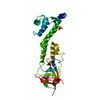
| Title | Crystal structure of the periplasmic domain of IgaA from Escherichia coli | ||||||
 Components Components | Intracellular growth attenuator protein igaA | ||||||
 Keywords Keywords | SIGNALING PROTEIN / Periplasmic protein / signal transduction / Structural Genomics / Center for Structural Biology of Infectious Diseases / CSBID / Center for Structural Genomics of Infectious Diseases / CSGID | ||||||
| Function / homology | Intracellular growth attenuator IgaA / Intracellular growth attenuator protein IgaA / plasma membrane / Intracellular growth attenuator protein igaA Function and homology information Function and homology information | ||||||
| Biological species |  | ||||||
| Method |  X-RAY DIFFRACTION / SYNCHROTRON / MOLECULAR REPLACEMENT / Resolution: 2.64 Å X-RAY DIFFRACTION / SYNCHROTRON / MOLECULAR REPLACEMENT / Resolution: 2.64 Å | ||||||
 Authors Authors | Watanabe, N. / Savchenko, A. / Center for Structural Biology of Infectious Diseases (CSBID) / Center for Structural Genomics of Infectious Diseases (CSGID) | ||||||
| Funding support |  United States, 1items United States, 1items
| ||||||
 Citation Citation | Journal: Structure / Year: 2024 Title: Molecular insights into the initiation step of the Rcs signaling pathway. Authors: Watanabe, N. / Savchenko, A. | ||||||
| History |
|
- Structure visualization
| Structure viewer | Molecule: MolmilJmol/JSmol |
|---|
- Downloads & links
-Download
| PDBx/mmCIF format | 9bj0.cif.gz | 122.4 KB | Display | PDBx/mmCIF format |
|---|---|---|---|---|
| PDB format | pdb9bj0.ent.gz | 86.1 KB | Display | PDB format |
| PDBx/mmJSON format | 9bj0.json.gz | Tree view | PDBx/mmJSON format | |
| Others |  Other downloads Other downloads |
-Validation report
| Arichive directory | https://data.pdbj.org/pub/pdb/validation_reports/bj/9bj0ftp://data.pdbj.org/pub/pdb/validation_reports/bj/9bj0 | HTTPS FTP |
|---|
-Related structure data

-Links
 PDBj
PDBj- Assembly
| Deposited unit | 
| ||||||||||||
|---|---|---|---|---|---|---|---|---|---|---|---|---|---|
| 1 | 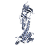
| ||||||||||||
| 2 | 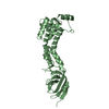
| ||||||||||||
| Unit cell |
| ||||||||||||
| Components on special symmetry positions |
|
-Components
| #1: Protein | Mass: 29556.217 Da / Num. of mol.: 2 / Fragment: periplasmic domain (UNP residues 203-475) Source method: isolated from a genetically manipulated source Source: (gene. exp.) #2: Water | ChemComp-HOH / |  Mass: 18.015 Da / Num. of mol.: 67 / Source method: isolated from a natural source / Formula: H2O Mass: 18.015 Da / Num. of mol.: 67 / Source method: isolated from a natural source / Formula: H2OHas protein modification | Y | |
|---|
-Experimental details
-Experiment
| Experiment | Method: X-RAY DIFFRACTION / Number of used crystals: 1 |
|---|
- Sample preparation
| Crystal | Density Matthews: 2.88 Å3/Da / Density % sol: 57.32 % |
|---|---|
| Crystal grow | Temperature: 294 K / Method: vapor diffusion / Details: 20% PEG3350, 0.2 M potassium citrate |
-Data collection
| Diffraction | Mean temperature: 100 K / Serial crystal experiment: N |
|---|---|
| Diffraction source | Source: SYNCHROTRON / Site: APS / Beamline: 19-ID / Wavelength: 0.97911 Å |
| Detector | Type: DECTRIS PILATUS 6M / Detector: PIXEL / Date: Nov 16, 2021 |
| Radiation | Protocol: SINGLE WAVELENGTH / Monochromatic (M) / Laue (L): M / Scattering type: x-ray |
| Radiation wavelength | Wavelength: 0.97911 Å / Relative weight: 1 |
| Reflection | Resolution: 2.64→40 Å / Num. obs: 20383 / % possible obs: 99.6 % / Redundancy: 6 % / Biso Wilson estimate: 74.17 Å2 / Rrim(I) all: 0.083 / Net I/σ(I): 29.5 |
| Reflection shell | Resolution: 2.65→2.74 Å / Num. unique obs: 1993 / CC1/2: 0.788 |
- Processing
| Software |
| |||||||||||||||||||||||||||||||||||||||||||||||||||||||||||||||
|---|---|---|---|---|---|---|---|---|---|---|---|---|---|---|---|---|---|---|---|---|---|---|---|---|---|---|---|---|---|---|---|---|---|---|---|---|---|---|---|---|---|---|---|---|---|---|---|---|---|---|---|---|---|---|---|---|---|---|---|---|---|---|---|---|
| Refinement | Method to determine structure: MOLECULAR REPLACEMENT / Resolution: 2.64→39.36 Å / SU ML: 0.3385 / Cross valid method: FREE R-VALUE / σ(F): 1.34 / Phase error: 29.7673 Stereochemistry target values: GeoStd + Monomer Library + CDL v1.2
| |||||||||||||||||||||||||||||||||||||||||||||||||||||||||||||||
| Solvent computation | Shrinkage radii: 0.9 Å / VDW probe radii: 1.11 Å / Solvent model: FLAT BULK SOLVENT MODEL | |||||||||||||||||||||||||||||||||||||||||||||||||||||||||||||||
| Displacement parameters | Biso mean: 81.24 Å2 | |||||||||||||||||||||||||||||||||||||||||||||||||||||||||||||||
| Refinement step | Cycle: LAST / Resolution: 2.64→39.36 Å
| |||||||||||||||||||||||||||||||||||||||||||||||||||||||||||||||
| Refine LS restraints |
| |||||||||||||||||||||||||||||||||||||||||||||||||||||||||||||||
| LS refinement shell |
|
