National Institutes of Health/National Institute of Neurological Disorders and Stroke (NIH/NINDS)
NS127292
米国
引用
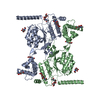
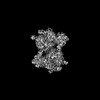
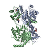
ジャーナル: J Biol Chem / 年: 2024 タイトル: Structure of a truncated human GlcNAc-1-phosphotransferase variant reveals the basis for its hyperactivity. 著者: Hua Li / Balraj Doray / Benjamin C Jennings / Wang-Sik Lee / Lin Liu / Stuart Kornfeld / Huilin Li / 要旨: Mutations that cause loss of function of GlcNAc-1-phosphotransferase (PTase) lead to the lysosomal storage disorder mucolipidosis II. PTase is the key enzyme of the mannose 6-phosphate (M6P) ...Mutations that cause loss of function of GlcNAc-1-phosphotransferase (PTase) lead to the lysosomal storage disorder mucolipidosis II. PTase is the key enzyme of the mannose 6-phosphate (M6P) targeting system that is responsible for tagging lysosomal hydrolases with the M6P moiety for their delivery to the lysosome. We had previously generated a truncated hyperactive form of PTase termed S1S3 which was shown to notably increase the phosphorylation level of secreted lysosomal enzymes and enhance their uptake by cells. Here, we report the 3.4 Å cryo-EM structure of soluble S1S3 lacking both transmembrane domains and cytosolic tails. The structure reveals a high degree of conservation of the catalytic core to full-length PTase. In this dimeric structure, the EF-hand of one protomer is observed interacting with the conserved region four of the other. In addition, we present a high-quality EM 3D map of the UDP-GlcNAc bound form of the full-length soluble protein showing the key molecular interactions between the nucleotide sugar donor and side chain amino acids of the protein. Finally, although the domain organization of S1S3 is very similar to that of the Drosophila melanogaster (fruit fly) PTase homolog, we establish that the latter does not act on lysosomal hydrolases.
履歴
登録
2024年4月18日
登録サイト: RCSB / 処理サイト: RCSB
改定 1.0
2025年2月26日
Provider: repository / タイプ: Initial release
改定 1.0
2025年2月26日
Data content type: EM metadata / Data content type: EM metadata / Provider: repository / タイプ: Initial release
改定 1.0
2025年2月26日
Data content type: FSC / Data content type: FSC / Provider: repository / タイプ: Initial release
改定 1.0
2025年2月26日
Data content type: Half map / Part number: 1 / Data content type: Half map / Provider: repository / タイプ: Initial release
改定 1.0
2025年2月26日
Data content type: Half map / Part number: 2 / Data content type: Half map / Provider: repository / タイプ: Initial release
改定 1.0
2025年2月26日
Data content type: Image / Data content type: Image / Provider: repository / タイプ: Initial release
改定 1.0
2025年2月26日
Data content type: Mask / Part number: 1 / Data content type: Mask / Provider: repository / タイプ: Initial release
改定 1.0
2025年2月26日
Data content type: Primary map / Data content type: Primary map / Provider: repository / タイプ: Initial release
 ムービー
ムービー コントローラー


 データを開く
 基本情報
基本情報 要素
要素 キーワード
キーワード 機能・相同性情報
 Homo sapiens (ヒト)
 データ登録者
 米国, 1件
  引用
引用 構造の表示
構造の表示 ダウンロードとリンク
 その他のダウンロード
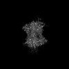
 PDBj
PDBj
 集合体
集合体

 Cricetulus griseus (モンゴルキヌゲネズミ)
 タイプ: D-saccharide, beta linking / 分子量: 221.208 Da / 分子数: 6 / 由来タイプ: 合成 / 式: C8H15NO6 / タイプ: SUBJECT OF INVESTIGATION


 分子量: 40.078 Da / 分子数: 2 / 由来タイプ: 合成 / 式: Ca / タイプ: SUBJECT OF INVESTIGATION
 分子量: 24.305 Da / 分子数: 4 / 由来タイプ: 合成 / 式: Mg / タイプ: SUBJECT OF INVESTIGATION
 分子量: 607.354 Da / 分子数: 2 / 由来タイプ: 合成 / 式: C17H27N3O17P2 / タイプ: SUBJECT OF INVESTIGATION
 試料調製
試料調製 電子顕微鏡撮影
 FIELD EMISSION GUN / 加速電圧: 300 kV / 照射モード: OTHER
 解析
解析