Movie
Movie Controller

[English] 日本語
 Yorodumi
- PDB-9bgf: Structure of human GlcNAc-1-phosphotransferase complexed with the... -
+ Open data

- Basic information
| Entry | Database: PDB / ID: 9bgf | |||||||||||||||||||||||||||
|---|---|---|---|---|---|---|---|---|---|---|---|---|---|---|---|---|---|---|---|---|---|---|---|---|---|---|---|---|
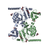
| Title | Structure of human GlcNAc-1-phosphotransferase complexed with the donor substrate UDP-GlcNAc | |||||||||||||||||||||||||||
 Components Components | N-acetylglucosamine-1-phosphotransferase subunits alpha/beta | |||||||||||||||||||||||||||
 Keywords Keywords | TRANSFERASE / GlcNAc-1-phosphotransferase / lysosomal hydrolases / mannose 6-phosphate trafficking pathway | |||||||||||||||||||||||||||
| Function / homology |  Function and homology information Function and homology informationUDP-N-acetylglucosamine-lysosomal-enzyme N-acetylglucosaminephosphotransferase / N-glycan processing to lysosome / UDP-N-acetylglucosamine-lysosomal-enzyme N-acetylglucosaminephosphotransferase activity / secretion of lysosomal enzymes / carbohydrate phosphorylation / lysosome organization / Golgi membrane / calcium ion binding / Golgi apparatus Similarity search - Function | |||||||||||||||||||||||||||
| Biological species |  Homo sapiens (human) Homo sapiens (human) | |||||||||||||||||||||||||||
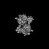
| Method | ELECTRON MICROSCOPY / single particle reconstruction / cryo EM / Resolution: 2.9 Å | |||||||||||||||||||||||||||
 Authors Authors | Li, H. / Li, H. | |||||||||||||||||||||||||||
| Funding support |  United States, 1items United States, 1items
| |||||||||||||||||||||||||||
 Citation Citation | Journal: J Biol Chem / Year: 2024 Title: Structure of a truncated human GlcNAc-1-phosphotransferase variant reveals the basis for its hyperactivity. Authors: Hua Li / Balraj Doray / Benjamin C Jennings / Wang-Sik Lee / Lin Liu / Stuart Kornfeld / Huilin Li / Abstract: Mutations that cause loss of function of GlcNAc-1-phosphotransferase (PTase) lead to the lysosomal storage disorder mucolipidosis II. PTase is the key enzyme of the mannose 6-phosphate (M6P) ...Mutations that cause loss of function of GlcNAc-1-phosphotransferase (PTase) lead to the lysosomal storage disorder mucolipidosis II. PTase is the key enzyme of the mannose 6-phosphate (M6P) targeting system that is responsible for tagging lysosomal hydrolases with the M6P moiety for their delivery to the lysosome. We had previously generated a truncated hyperactive form of PTase termed S1S3 which was shown to notably increase the phosphorylation level of secreted lysosomal enzymes and enhance their uptake by cells. Here, we report the 3.4 Å cryo-EM structure of soluble S1S3 lacking both transmembrane domains and cytosolic tails. The structure reveals a high degree of conservation of the catalytic core to full-length PTase. In this dimeric structure, the EF-hand of one protomer is observed interacting with the conserved region four of the other. In addition, we present a high-quality EM 3D map of the UDP-GlcNAc bound form of the full-length soluble protein showing the key molecular interactions between the nucleotide sugar donor and side chain amino acids of the protein. Finally, although the domain organization of S1S3 is very similar to that of the Drosophila melanogaster (fruit fly) PTase homolog, we establish that the latter does not act on lysosomal hydrolases. | |||||||||||||||||||||||||||
| History |
|
- Structure visualization
| Structure viewer | Molecule: MolmilJmol/JSmol |
|---|
- Downloads & links
-Download
| PDBx/mmCIF format | 9bgf.cif.gz | 226.3 KB | Display | PDBx/mmCIF format |
|---|---|---|---|---|
| PDB format | pdb9bgf.ent.gz | 158.4 KB | Display | PDB format |
| PDBx/mmJSON format | 9bgf.json.gz | Tree view | PDBx/mmJSON format | |
| Others |  Other downloads Other downloads |
-Validation report
| Arichive directory | https://data.pdbj.org/pub/pdb/validation_reports/bg/9bgfftp://data.pdbj.org/pub/pdb/validation_reports/bg/9bgf | HTTPS FTP |
|---|
-Related structure data
| Related structure data | 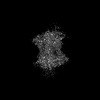 44511MC 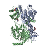 9bggC M: map data used to model this data C: citing same article ( |
|---|---|
| Similar structure data |
-Links
 PDBj
PDBj
- Assembly
| Deposited unit | 
|
|---|---|
| 1 |
|
-Components
-Protein , 1 types, 2 molecules AB
| #1: Protein | Mass: 134787.500 Da / Num. of mol.: 2 / Fragment: UNP residues 44-1209 Source method: isolated from a genetically manipulated source Source: (gene. exp.) Homo sapiens (human) / Gene: GNPTAB, GNPTA, KIAA1208 / Production host:   Cricetulus griseus (Chinese hamster) Cricetulus griseus (Chinese hamster)References: UniProt: Q3T906, UDP-N-acetylglucosamine-lysosomal-enzyme N-acetylglucosaminephosphotransferase |
|---|
-Sugars , 2 types, 7 molecules 
| #2: Polysaccharide | 2-acetamido-2-deoxy-beta-D-glucopyranose-(1-4)-2-acetamido-2-deoxy-beta-D-glucopyranose Source method: isolated from a genetically manipulated source |
|---|---|
| #6: Sugar | ChemComp-NAG /  Type: D-saccharide, beta linking / Mass: 221.208 Da / Num. of mol.: 6 / Source method: obtained synthetically / Formula: C8H15NO6 / Feature type: SUBJECT OF INVESTIGATION Type: D-saccharide, beta linking / Mass: 221.208 Da / Num. of mol.: 6 / Source method: obtained synthetically / Formula: C8H15NO6 / Feature type: SUBJECT OF INVESTIGATION |
-Non-polymers , 3 types, 8 molecules 


| #3: Chemical |  Mass: 40.078 Da / Num. of mol.: 2 / Source method: obtained synthetically / Formula: Ca / Feature type: SUBJECT OF INVESTIGATION Mass: 40.078 Da / Num. of mol.: 2 / Source method: obtained synthetically / Formula: Ca / Feature type: SUBJECT OF INVESTIGATION#4: Chemical | ChemComp-MG /  Mass: 24.305 Da / Num. of mol.: 4 / Source method: obtained synthetically / Formula: Mg / Feature type: SUBJECT OF INVESTIGATION Mass: 24.305 Da / Num. of mol.: 4 / Source method: obtained synthetically / Formula: Mg / Feature type: SUBJECT OF INVESTIGATION#5: Chemical |  Mass: 607.354 Da / Num. of mol.: 2 / Source method: obtained synthetically / Formula: C17H27N3O17P2 / Feature type: SUBJECT OF INVESTIGATION Mass: 607.354 Da / Num. of mol.: 2 / Source method: obtained synthetically / Formula: C17H27N3O17P2 / Feature type: SUBJECT OF INVESTIGATION |
|---|
-Details
| Has ligand of interest | Y |
|---|---|
| Has protein modification | Y |
-Experimental details
-Experiment
| Experiment | Method: ELECTRON MICROSCOPY |
|---|---|
| EM experiment | Aggregation state: PARTICLE / 3D reconstruction method: single particle reconstruction |
- Sample preparation
| Component | Name: GlcNAc-1-phosphotransferase / Type: COMPLEX / Entity ID: #1 / Source: RECOMBINANT | ||||||||||||||||||||
|---|---|---|---|---|---|---|---|---|---|---|---|---|---|---|---|---|---|---|---|---|---|
| Molecular weight | Value: 0.268 MDa / Experimental value: YES | ||||||||||||||||||||
| Source (natural) | Organism: Homo sapiens (human) | ||||||||||||||||||||
| Source (recombinant) | Organism: Cricetulus griseus (Chinese hamster) | ||||||||||||||||||||
| Buffer solution | pH: 7.8 | ||||||||||||||||||||
| Buffer component |
| ||||||||||||||||||||
| Specimen | Conc.: 0.15 mg/ml / Embedding applied: NO / Shadowing applied: NO / Staining applied: NO / Vitrification applied: YES | ||||||||||||||||||||
| Specimen support | Grid material: COPPER / Grid mesh size: 300 divisions/in. / Grid type: Quantifoil R2/1 | ||||||||||||||||||||
| Vitrification | Instrument: FEI VITROBOT MARK IV / Cryogen name: ETHANE / Humidity: 95 % / Chamber temperature: 299 K |
- Electron microscopy imaging
| Experimental equipment |  Model: Titan Krios / Image courtesy: FEI Company |
|---|---|
| Microscopy | Model: FEI TITAN KRIOS |
| Electron gun | Electron source:  FIELD EMISSION GUN / Accelerating voltage: 300 kV / Illumination mode: OTHER FIELD EMISSION GUN / Accelerating voltage: 300 kV / Illumination mode: OTHER |
| Electron lens | Mode: BRIGHT FIELD / Nominal magnification: 105000 X / Nominal defocus max: 1600 nm / Nominal defocus min: 1000 nm / Cs: 2.7 mm / C2 aperture diameter: 70 µm / Alignment procedure: COMA FREE |
| Specimen holder | Cryogen: NITROGEN / Specimen holder model: FEI TITAN KRIOS AUTOGRID HOLDER / Temperature (max): 193 K / Temperature (min): 193 K / Residual tilt: 0.05 mradians |
| Image recording | Average exposure time: 1 sec. / Electron dose: 60 e/Å2 / Film or detector model: GATAN K3 (6k x 4k) / Num. of grids imaged: 1 / Num. of real images: 24130 |
| Image scans | Width: 5760 / Height: 4092 |
- Processing
| EM software | Name: PHENIX / Version: 1.21.1_5286: / Category: model refinement | ||||||||||||||||||||||||
|---|---|---|---|---|---|---|---|---|---|---|---|---|---|---|---|---|---|---|---|---|---|---|---|---|---|
| CTF correction | Type: PHASE FLIPPING AND AMPLITUDE CORRECTION | ||||||||||||||||||||||||
| Particle selection | Num. of particles selected: 2859876 | ||||||||||||||||||||||||
| Symmetry | Point symmetry: C1 (asymmetric) | ||||||||||||||||||||||||
| 3D reconstruction | Resolution: 2.9 Å / Resolution method: FSC 0.143 CUT-OFF / Num. of particles: 876204 / Symmetry type: POINT | ||||||||||||||||||||||||
| Atomic model building | PDB-ID: 7s05 Accession code: 7s05 / Source name: PDB / Type: experimental model | ||||||||||||||||||||||||
| Refine LS restraints |
|