 Movie
Movie Controller

[English] 日本語
 Yorodumi
- PDB-9bey: Structure of a lambda-carrageenan active GH2 A in complex with in... -
+ Open data

- Basic information
| Entry | Database: PDB / ID: 9bey | ||||||
|---|---|---|---|---|---|---|---|
| Title | Structure of a lambda-carrageenan active GH2 A in complex with inhibitor | ||||||
 Components Components | GH2 beta-galactosidase | ||||||
 Keywords Keywords | SUGAR BINDING PROTEIN / Sulfatase / carrageenan | ||||||
| Function / homology | D-galacto-isofagomine Function and homology information Function and homology information | ||||||
| Biological species |  Pseudoalteromonas distincta (bacteria) Pseudoalteromonas distincta (bacteria) | ||||||
| Method |  X-RAY DIFFRACTION / MOLECULAR REPLACEMENT / Resolution: 2.386 Å X-RAY DIFFRACTION / MOLECULAR REPLACEMENT / Resolution: 2.386 Å | ||||||
 Authors Authors | Hettle, J.A. / Vickers, C. / Boraston, A.B. | ||||||
| Funding support |  Canada, 1items Canada, 1items
| ||||||
 Citation Citation | Journal: To Be Published Title: Structure of a lambda-carrageenan active GH2 A in complex with inhibitor Authors: Hettle, J.A. / Vickers, C. / Boraston, A.B. | ||||||
| History |
|
- Structure visualization
| Structure viewer | Molecule: MolmilJmol/JSmol |
|---|
- Downloads & links
-Download
| PDBx/mmCIF format | 9bey.cif.gz | 180.6 KB | Display | PDBx/mmCIF format |
|---|---|---|---|---|
| PDB format | pdb9bey.ent.gz | 139.1 KB | Display | PDB format |
| PDBx/mmJSON format | 9bey.json.gz | Tree view | PDBx/mmJSON format | |
| Others |  Other downloads Other downloads |
-Validation report
| Arichive directory | https://data.pdbj.org/pub/pdb/validation_reports/be/9beyftp://data.pdbj.org/pub/pdb/validation_reports/be/9bey | HTTPS FTP |
|---|
-Related structure data
| Similar structure data |
|---|
-Links
 PDBj
PDBj- Assembly
| Deposited unit | 
| ||||||||
|---|---|---|---|---|---|---|---|---|---|
| 1 |
| ||||||||
| Unit cell |
|
-Components
| #1: Protein | Mass: 93714.508 Da / Num. of mol.: 1 Source method: isolated from a genetically manipulated source Source: (gene. exp.) Pseudoalteromonas distincta (bacteria) / Production host: | ||||||
|---|---|---|---|---|---|---|---|
| #2: Chemical | ChemComp-GIF / 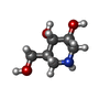  Mass: 147.172 Da / Num. of mol.: 1 / Source method: obtained synthetically / Formula: C6H13NO3 / Feature type: SUBJECT OF INVESTIGATION Mass: 147.172 Da / Num. of mol.: 1 / Source method: obtained synthetically / Formula: C6H13NO3 / Feature type: SUBJECT OF INVESTIGATION | ||||||
| #3: Chemical | ChemComp-EDO /   Mass: 62.068 Da / Num. of mol.: 12 / Source method: obtained synthetically / Formula: C2H6O2 Mass: 62.068 Da / Num. of mol.: 12 / Source method: obtained synthetically / Formula: C2H6O2#4: Water | ChemComp-HOH / |  Mass: 18.015 Da / Num. of mol.: 393 / Source method: isolated from a natural source / Formula: H2O Mass: 18.015 Da / Num. of mol.: 393 / Source method: isolated from a natural source / Formula: H2OHas ligand of interest | Y | Has protein modification | N | |
-Experimental details
-Experiment
| Experiment | Method: X-RAY DIFFRACTION / Number of used crystals: 1 |
|---|
- Sample preparation
| Crystal | Density Matthews: 2.65 Å3/Da / Density % sol: 53.61 % |
|---|---|
| Crystal grow | Temperature: 291 K / Method: vapor diffusion, hanging drop / pH: 6.5 Details: 15% PEG 8000, 0.1 M sodium cacodylate, 0.2 M calcium acetate, 0.2 M LiCl, 5% glycerol |
-Data collection
| Diffraction | Mean temperature: 100 K / Serial crystal experiment: N |
|---|---|
| Diffraction source | Source: ROTATING ANODE / Type: RIGAKU MICROMAX-007 HF / Wavelength: 1.541 Å |
| Detector | Type: DECTRIS PILATUS 200K / Detector: PIXEL / Date: Apr 14, 2020 |
| Radiation | Protocol: SINGLE WAVELENGTH / Monochromatic (M) / Laue (L): M / Scattering type: x-ray |
| Radiation wavelength | Wavelength: 1.541 Å / Relative weight: 1 |
| Reflection | Resolution: 2.386→30 Å / Num. obs: 38258 / % possible obs: 97.9 % / Redundancy: 3.7 % / CC1/2: 0.987 / Rmerge(I) obs: 0.121 / Rpim(I) all: 0.058 / Net I/σ(I): 11.3 |
| Reflection shell | Resolution: 2.386→2.44 Å / Rmerge(I) obs: 0.318 / Num. unique obs: 1529 / CC1/2: 0.821 / Rpim(I) all: 0.231 |
- Processing
| Software |
| |||||||||||||||||||||||||||||||||||||||||||||||||||||||||||||||||||||||||||||||||||||||||||||||||||||||||
|---|---|---|---|---|---|---|---|---|---|---|---|---|---|---|---|---|---|---|---|---|---|---|---|---|---|---|---|---|---|---|---|---|---|---|---|---|---|---|---|---|---|---|---|---|---|---|---|---|---|---|---|---|---|---|---|---|---|---|---|---|---|---|---|---|---|---|---|---|---|---|---|---|---|---|---|---|---|---|---|---|---|---|---|---|---|---|---|---|---|---|---|---|---|---|---|---|---|---|---|---|---|---|---|---|---|---|
| Refinement | Method to determine structure: MOLECULAR REPLACEMENT / Resolution: 2.386→29.676 Å / SU ML: 0.3 / Cross valid method: FREE R-VALUE / σ(F): 1.34 / Phase error: 23.12 / Stereochemistry target values: ML
| |||||||||||||||||||||||||||||||||||||||||||||||||||||||||||||||||||||||||||||||||||||||||||||||||||||||||
| Solvent computation | Shrinkage radii: 0.9 Å / VDW probe radii: 1.11 Å / Solvent model: FLAT BULK SOLVENT MODEL | |||||||||||||||||||||||||||||||||||||||||||||||||||||||||||||||||||||||||||||||||||||||||||||||||||||||||
| Refinement step | Cycle: LAST / Resolution: 2.386→29.676 Å
| |||||||||||||||||||||||||||||||||||||||||||||||||||||||||||||||||||||||||||||||||||||||||||||||||||||||||
| Refine LS restraints |
| |||||||||||||||||||||||||||||||||||||||||||||||||||||||||||||||||||||||||||||||||||||||||||||||||||||||||
| LS refinement shell |
|
