 Movie
Movie Controller

[English] 日本語
 Yorodumi
- PDB-9bc8: Cargo-loaded Myxococcus xanthus EncA encapsulin engineered pore m... -
+ Open data

- Basic information
| Entry | Database: PDB / ID: 9bc8 | ||||||
|---|---|---|---|---|---|---|---|
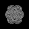
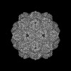
| Title | Cargo-loaded Myxococcus xanthus EncA encapsulin engineered pore mutant with T=4 icosahedral symmetry | ||||||
 Components Components |
| ||||||
 Keywords Keywords | VIRUS LIKE PARTICLE / encapsulin / nanocompartment / pore mutant | ||||||
| Function / homology |  Function and homology information Function and homology informationencapsulin nanocompartment / iron ion transport / intracellular iron ion homeostasis / metal ion binding Similarity search - Function | ||||||
| Biological species |  Myxococcus xanthus DK 1622 (bacteria) Myxococcus xanthus DK 1622 (bacteria) | ||||||
| Method | ELECTRON MICROSCOPY / single particle reconstruction / cryo EM / Resolution: 3.46 Å | ||||||
 Authors Authors | Andreas, M.P. / Kwon, S. / Giessen, T.W. | ||||||
| Funding support |  United States, 1items United States, 1items
| ||||||
 Citation Citation | Journal: ACS Nano / Year: 2024 Title: Pore Engineering as a General Strategy to Improve Protein-Based Enzyme Nanoreactor Performance. Authors: Seokmu Kwon / Michael P Andreas / Tobias W Giessen / Abstract: Enzyme nanoreactors are nanoscale compartments consisting of encapsulated enzymes and a selectively permeable barrier. Sequestration and colocalization of enzymes can increase catalytic activity, ...Enzyme nanoreactors are nanoscale compartments consisting of encapsulated enzymes and a selectively permeable barrier. Sequestration and colocalization of enzymes can increase catalytic activity, stability, and longevity, highly desirable features for many biotechnological and biomedical applications of enzyme catalysts. One promising strategy to construct enzyme nanoreactors is to repurpose protein nanocages found in nature. However, protein-based enzyme nanoreactors often exhibit decreased catalytic activity, partially caused by a mismatch of protein shell selectivity and the substrate requirements of encapsulated enzymes. No broadly applicable and modular protein-based nanoreactor platform is currently available. Here, we introduce a pore-engineered universal enzyme nanoreactor platform based on encapsulins-microbial self-assembling protein nanocompartments with programmable and selective enzyme packaging capabilities. We structurally characterize our protein shell designs via cryo-electron microscopy and highlight their polymorphic nature. Through fluorescence polarization assays, we show their improved molecular flux behavior and highlight their expanded substrate range via a number of proof-of-concept enzyme nanoreactor designs. This work lays the foundation for utilizing our encapsulin-based nanoreactor platform for diverse future biotechnological and biomedical applications. | ||||||
| History |
|
- Structure visualization
| Structure viewer | Molecule: MolmilJmol/JSmol |
|---|
- Downloads & links
-Download
| PDBx/mmCIF format | 9bc8.cif.gz | 222.9 KB | Display | PDBx/mmCIF format |
|---|---|---|---|---|
| PDB format | pdb9bc8.ent.gz | 181.3 KB | Display | PDB format |
| PDBx/mmJSON format | 9bc8.json.gz | Tree view | PDBx/mmJSON format | |
| Others |  Other downloads Other downloads |
-Validation report
| Arichive directory | https://data.pdbj.org/pub/pdb/validation_reports/bc/9bc8ftp://data.pdbj.org/pub/pdb/validation_reports/bc/9bc8 | HTTPS FTP |
|---|
-Related structure data
| Related structure data |  44427MC 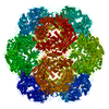 9b9iC 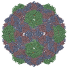 9b9qC M: map data used to model this data C: citing same article ( |
|---|---|
| Similar structure data |
-Links
 PDBj
PDBj
- Assembly
| Deposited unit | 
|
|---|---|
| 1 | x 60
|
| 2 |
|
| 3 | x 5
|
| 4 | x 6
|
| 5 | 
|
| Symmetry | Point symmetry: (Schoenflies symbol: I (icosahedral)) |
-Components
| #1: Protein | Mass: 30901.014 Da / Num. of mol.: 4 / Mutation: I203G, Y204G Source method: isolated from a genetically manipulated source Details: del(205-210) / Source: (gene. exp.) Myxococcus xanthus DK 1622 (bacteria) / Gene: encA, MXAN_3556 / Production host: #2: Protein/peptide | Mass: 1415.685 Da / Num. of mol.: 4 Fragment: C-terminal targeting peptide (UNP residues 119-130) Source method: isolated from a genetically manipulated source Details: C-terminal targeting peptide of a SNAP-Tag-targeting peptide cargo protein. Source: (gene. exp.) Myxococcus xanthus DK 1622 (bacteria) / Gene: encC, MXAN_4464 / Production host: Has protein modification | N | |
|---|
-Experimental details
-Experiment
| Experiment | Method: ELECTRON MICROSCOPY |
|---|---|
| EM experiment | Aggregation state: PARTICLE / 3D reconstruction method: single particle reconstruction |
- Sample preparation
| Component |
| ||||||||||||||||||||||||
|---|---|---|---|---|---|---|---|---|---|---|---|---|---|---|---|---|---|---|---|---|---|---|---|---|---|
| Molecular weight |
| ||||||||||||||||||||||||
| Source (natural) |
| ||||||||||||||||||||||||
| Source (recombinant) |
| ||||||||||||||||||||||||
| Buffer solution | pH: 7.5 / Details: 150 mM NaCl, 20 mM Tris pH 7.5 | ||||||||||||||||||||||||
| Buffer component |
| ||||||||||||||||||||||||
| Specimen | Conc.: 4.1 mg/ml / Embedding applied: NO / Shadowing applied: NO / Staining applied: NO / Vitrification applied: YES | ||||||||||||||||||||||||
| Specimen support | Details: Glow discharge was performed under vacuum at 5 mA for 60 seconds. Grid material: COPPER / Grid mesh size: 200 divisions/in. / Grid type: Quantifoil R1.2/1.3 | ||||||||||||||||||||||||
| Vitrification | Instrument: FEI VITROBOT MARK IV / Cryogen name: ETHANE / Humidity: 100 % / Chamber temperature: 295 K Details: Blot force: 20 Blot time: 4 seconds Wait time: 0 seconds |
- Electron microscopy imaging
| Experimental equipment |  Model: Talos Arctica / Image courtesy: FEI Company |
|---|---|
| Microscopy | Model: FEI TECNAI ARCTICA |
| Electron gun | Electron source:  FIELD EMISSION GUN / Accelerating voltage: 200 kV / Illumination mode: FLOOD BEAM FIELD EMISSION GUN / Accelerating voltage: 200 kV / Illumination mode: FLOOD BEAM |
| Electron lens | Mode: BRIGHT FIELD / Nominal magnification: 45000 X / Nominal defocus max: 1800 nm / Nominal defocus min: 1000 nm |
| Image recording | Average exposure time: 6 sec. / Electron dose: 39.56 e/Å2 / Detector mode: COUNTING / Film or detector model: GATAN K2 SUMMIT (4k x 4k) / Num. of grids imaged: 1 / Num. of real images: 706 |
| Image scans | Width: 3710 / Height: 3838 / Movie frames/image: 30 |
- Processing
| EM software |
| ||||||||||||||||||||||||||||||||||||||||||||||||||
|---|---|---|---|---|---|---|---|---|---|---|---|---|---|---|---|---|---|---|---|---|---|---|---|---|---|---|---|---|---|---|---|---|---|---|---|---|---|---|---|---|---|---|---|---|---|---|---|---|---|---|---|
| CTF correction | Type: PHASE FLIPPING AND AMPLITUDE CORRECTION | ||||||||||||||||||||||||||||||||||||||||||||||||||
| Particle selection | Num. of particles selected: 37600 | ||||||||||||||||||||||||||||||||||||||||||||||||||
| Symmetry | Point symmetry: I (icosahedral) | ||||||||||||||||||||||||||||||||||||||||||||||||||
| 3D reconstruction | Resolution: 3.46 Å / Resolution method: FSC 0.143 CUT-OFF / Num. of particles: 4386 Details: Homogeneous refinement was performed against the intial ab-initio map using I symmetry, per-particle defocus optimization, per-group CTF parameterization, and Ewald sphere correction. Symmetry type: POINT | ||||||||||||||||||||||||||||||||||||||||||||||||||
| Atomic model building | B value: 60.6 / Protocol: FLEXIBLE FIT / Space: REAL / Target criteria: cross-correlation coefficient Details: ChimeraX v1.2.5 was first used to place the starting model (PDB: 7S4Q) in the cryo-EM map by using the fit in map command. The model was then manually refined using Coot v 0.9.8.1, followed ...Details: ChimeraX v1.2.5 was first used to place the starting model (PDB: 7S4Q) in the cryo-EM map by using the fit in map command. The model was then manually refined using Coot v 0.9.8.1, followed by iterative real-space refinements in Phenix v1.20.1-4487-000. BioMT operators were identified from the cryo-EM map using map_symmetry command in Phenix then applied to the model using the apply_ncs command to assemble the complete shell. Real-space refinement was repeated in Phenix with NCS constraints applied. The BioMT operators were then identified using find_ncs command in Phenix and applied to the header of a protomer of the NCS-refined model. | ||||||||||||||||||||||||||||||||||||||||||||||||||
| Atomic model building | PDB-ID: 7S4Q Accession code: 7S4Q / Source name: PDB / Type: experimental model |

