[English] 日本語
Yorodumi
- PDB-9bbh: Co-crystal structure of human DDB1 bound to fragment UB028670 -

+
Open data


ID or keywords:

Loading...

-
Basic information

Entry
Database: PDB / ID: 9bbh
TitleCo-crystal structure of human DDB1 bound to fragment UB028670
ComponentsDNA damage-binding protein 1
KeywordsLIGASE / WD-repeat / WDR / DDB1 / SGC
Function / homology
Function and homology information


positive regulation by virus of viral protein levels in host cell / epigenetic programming in the zygotic pronuclei / spindle assembly involved in female meiosis / Cul4-RING E3 ubiquitin ligase complex / UV-damage excision repair / biological process involved in interaction with symbiont / WD40-repeat domain binding / regulation of mitotic cell cycle phase transition / Cul4A-RING E3 ubiquitin ligase complex / Cul4B-RING E3 ubiquitin ligase complex ...positive regulation by virus of viral protein levels in host cell / epigenetic programming in the zygotic pronuclei / spindle assembly involved in female meiosis / Cul4-RING E3 ubiquitin ligase complex / UV-damage excision repair / biological process involved in interaction with symbiont / WD40-repeat domain binding / regulation of mitotic cell cycle phase transition / Cul4A-RING E3 ubiquitin ligase complex / Cul4B-RING E3 ubiquitin ligase complex / ubiquitin ligase complex scaffold activity / negative regulation of reproductive process / negative regulation of developmental process / cullin family protein binding / viral release from host cell / ectopic germ cell programmed cell death / proteasomal protein catabolic process / positive regulation of viral genome replication / positive regulation of gluconeogenesis / nucleotide-excision repair / Recognition of DNA damage by PCNA-containing replication complex / DNA Damage Recognition in GG-NER / regulation of circadian rhythm / Dual Incision in GG-NER / Transcription-Coupled Nucleotide Excision Repair (TC-NER) / Formation of TC-NER Pre-Incision Complex / Wnt signaling pathway / Formation of Incision Complex in GG-NER / Dual incision in TC-NER / Gap-filling DNA repair synthesis and ligation in TC-NER / positive regulation of protein catabolic process / cellular response to UV / rhythmic process / protein-macromolecule adaptor activity / Neddylation / site of double-strand break / ubiquitin-dependent protein catabolic process / proteasome-mediated ubiquitin-dependent protein catabolic process / chromosome, telomeric region / damaged DNA binding / protein ubiquitination / DNA repair / apoptotic process / DNA damage response / protein-containing complex binding / nucleolus / negative regulation of apoptotic process / protein-containing complex / DNA binding / extracellular space / extracellular exosome / nucleoplasm / nucleus / cytoplasm
Similarity search - Function
Cleavage/polyadenylation specificity factor, A subunit, N-terminal / Mono-functional DNA-alkylating methyl methanesulfonate N-term / Cleavage/polyadenylation specificity factor, A subunit, C-terminal / CPSF A subunit region / WD40-repeat-containing domain superfamily / WD40/YVTN repeat-like-containing domain superfamily
Similarity search - Domain/homology
L(+)-TARTARIC ACID / 4-methoxy-1H-indole / DNA damage-binding protein 1
Similarity search - Component
Biological speciesHomo sapiens (human)
MethodX-RAY DIFFRACTION / MOLECULAR REPLACEMENT / Resolution: 2 Å
AuthorsZeng, H. / Dong, A. / Frommlet, A. / Seitova, A. / Loppnau, P. / Ackloo, S. / Arrowsmith, C.H. / Edwards, A.M. / Halabelian, L. / Structural Genomics Consortium (SGC)
Funding support1items
OrganizationGrant numberCountry
Other private
CitationJournal: To be published
Title: Co-crystal structure of human DDB1 bound to fragment UB028670
Authors: Zeng, H. / Dong, A. / Frommlet, A. / Seitova, A. / Loppnau, P. / Ackloo, S. / Arrowsmith, C.H. / Edwards, A.M. / Halabelian, L. / Structural Genomics Consortium (SGC)
History
DepositionApr 5, 2024Deposition site: RCSB / Processing site: RCSB
Revision 1.0Jun 12, 2024Provider: repository / Type: Initial release

-
Structure visualization

Structure viewerMolecule:
MolmilJmol/JSmol

Downloads & links

-
Assembly

Deposited unit
A: DNA damage-binding protein 1
hetero molecules


Theoretical massNumber of molelcules
Total (without water)127,96012
Polymers127,2421
Non-polymers71911
Water11,566642
1


  • Idetical with deposited unit
  • defined by author
TypeNameSymmetry operationNumber
identity operation1_555x,y,z1
Unit cell
Length a, b, c (Å)62.924, 125.268, 168.529
Angle α, β, γ (deg.)90.00, 90.00, 90.00
Int Tables number19
Space group name H-MP212121

-
Components

-
Protein , 1 types, 1 molecules A

#1: Protein DNA damage-binding protein 1 / DDB p127 subunit / DNA damage-binding protein a / DDBa / Damage-specific DNA-binding protein 1 / ...DDB p127 subunit / DNA damage-binding protein a / DDBa / Damage-specific DNA-binding protein 1 / HBV X-associated protein 1 / XAP-1 / UV-damaged DNA-binding factor / UV-damaged DNA-binding protein 1 / UV-DDB 1 / XPE-binding factor / XPE-BF / Xeroderma pigmentosum group E-complementing protein / XPCe


Mass: 127241.594 Da / Num. of mol.: 1
Source method: isolated from a genetically manipulated source
Source: (gene. exp.) Homo sapiens (human) / Gene: DDB1, XAP1 / Production host: 
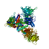
Spodoptera frugiperda (fall armyworm) / References: UniProt: Q16531

-
Non-polymers , 5 types, 653 molecules

#2: Chemical ChemComp-TLA / L(+)-TARTARIC ACID


Mass: 150.087 Da / Num. of mol.: 2 / Source method: obtained synthetically / Formula: C4H6O6
#3: Chemical ChemComp-VVP / 4-methoxy-1H-indole


Mass: 147.174 Da / Num. of mol.: 2 / Source method: isolated from a natural source / Formula: C9H9NO / Feature type: SUBJECT OF INVESTIGATION
#4: Chemical ChemComp-EDO / 1,2-ETHANEDIOL / ETHYLENE GLYCOL


Mass: 62.068 Da / Num. of mol.: 2 / Source method: obtained synthetically / Formula: C2H6O2
#5: Chemical
ChemComp-UNX / UNKNOWN ATOM OR ION


Num. of mol.: 5 / Source method: obtained synthetically
#6: Water ChemComp-HOH / water


Mass: 18.015 Da / Num. of mol.: 642 / Source method: isolated from a natural source / Formula: H2O

-
Details

Has ligand of interestY

-
Experimental details

-
Experiment

ExperimentMethod: X-RAY DIFFRACTION / Number of used crystals: 1

-
Sample preparation

CrystalDensity Matthews: 2.61 Å3/Da / Density % sol: 52.87 %
Crystal growTemperature: 291 K / Method: vapor diffusion, sitting drop / Details: 20% (v/w) PEG 3350, 0.2 M di-NH4tart

-
Data collection

DiffractionMean temperature: 100 K / Serial crystal experiment: N
Diffraction sourceSource: ROTATING ANODE / Type: RIGAKU FR-E SUPERBRIGHT / Wavelength: 1.54 Å
DetectorType: RIGAKU SATURN A200 / Detector: CCD / Date: Sep 14, 2021
RadiationProtocol: SINGLE WAVELENGTH / Monochromatic (M) / Laue (L): M / Scattering type: x-ray
Radiation wavelengthWavelength: 1.54 Å / Relative weight: 1
ReflectionResolution: 2→46.82 Å / Num. obs: 90240 / % possible obs: 99.4 % / Redundancy: 8 % / CC1/2: 0.999 / Rmerge(I) obs: 0.11 / Rpim(I) all: 0.041 / Rrim(I) all: 0.117 / Χ2: 0.99 / Net I/σ(I): 16.7
Reflection shellResolution: 2→2.03 Å / % possible obs: 97.3 % / Redundancy: 8 % / Rmerge(I) obs: 0.819 / Num. measured all: 34490 / Num. unique obs: 4336 / CC1/2: 0.751 / Rpim(I) all: 0.305 / Rrim(I) all: 0.875 / Χ2: 1.01 / Net I/σ(I) obs: 2.7

-
Processing

Software
NameVersionClassification
REFMAC5.8.0425refinement
Aimlessdata scaling
PHASERphasing
RefinementMethod to determine structure: MOLECULAR REPLACEMENT / Resolution: 2→46.82 Å / Cor.coef. Fo:Fc: 0.95 / Cor.coef. Fo:Fc free: 0.926 / SU B: 3.948 / SU ML: 0.108 / Cross valid method: THROUGHOUT / ESU R: 0.168 / ESU R Free: 0.156 / Stereochemistry target values: MAXIMUM LIKELIHOOD / Details: HYDROGENS HAVE BEEN ADDED IN THE RIDING POSITIONS
RfactorNum. reflection% reflectionSelection details
Rfree0.23191 4384 4.9 %RANDOM
Rwork0.1878 ---
obs0.18992 85778 99.3 %-
Solvent computationIon probe radii: 0.8 Å / Shrinkage radii: 0.8 Å / VDW probe radii: 1.2 Å / Solvent model: MASK
Displacement parametersBiso mean: 25.432 Å2
Baniso -1Baniso -2Baniso -3
1-0.64 Å2-0 Å20 Å2
2---0.62 Å2-0 Å2
3----0.01 Å2
Refinement stepCycle: 1 / Resolution: 2→46.82 Å
ProteinNucleic acidLigandSolventTotal
Num. atoms8497 0 55 642 9194
Refine LS restraints
Refine-IDTypeDev idealDev ideal targetNumber
X-RAY DIFFRACTIONr_bond_refined_d0.0070.0128846
X-RAY DIFFRACTIONr_bond_other_d0.0010.0168334
X-RAY DIFFRACTIONr_angle_refined_deg1.1351.81712029
X-RAY DIFFRACTIONr_angle_other_deg0.3881.7419167
X-RAY DIFFRACTIONr_dihedral_angle_1_deg8.02651130
X-RAY DIFFRACTIONr_dihedral_angle_2_deg7.372545
X-RAY DIFFRACTIONr_dihedral_angle_3_deg13.349101476
X-RAY DIFFRACTIONr_dihedral_angle_4_deg
X-RAY DIFFRACTIONr_chiral_restr0.0560.21400
X-RAY DIFFRACTIONr_gen_planes_refined0.0080.0210488
X-RAY DIFFRACTIONr_gen_planes_other0.0010.021966
X-RAY DIFFRACTIONr_nbd_refined
X-RAY DIFFRACTIONr_nbd_other
X-RAY DIFFRACTIONr_nbtor_refined
X-RAY DIFFRACTIONr_nbtor_other
X-RAY DIFFRACTIONr_xyhbond_nbd_refined
X-RAY DIFFRACTIONr_xyhbond_nbd_other
X-RAY DIFFRACTIONr_metal_ion_refined
X-RAY DIFFRACTIONr_metal_ion_other
X-RAY DIFFRACTIONr_symmetry_vdw_refined
X-RAY DIFFRACTIONr_symmetry_vdw_other
X-RAY DIFFRACTIONr_symmetry_hbond_refined
X-RAY DIFFRACTIONr_symmetry_hbond_other
X-RAY DIFFRACTIONr_symmetry_metal_ion_refined
X-RAY DIFFRACTIONr_symmetry_metal_ion_other
X-RAY DIFFRACTIONr_mcbond_it2.3562.6434490
X-RAY DIFFRACTIONr_mcbond_other2.3532.6434490
X-RAY DIFFRACTIONr_mcangle_it3.5734.7235630
X-RAY DIFFRACTIONr_mcangle_other3.5734.7235631
X-RAY DIFFRACTIONr_scbond_it2.9942.9264356
X-RAY DIFFRACTIONr_scbond_other2.9942.9274357
X-RAY DIFFRACTIONr_scangle_it
X-RAY DIFFRACTIONr_scangle_other4.7385.2566400
X-RAY DIFFRACTIONr_long_range_B_refined6.5525.159219
X-RAY DIFFRACTIONr_long_range_B_other6.50324.79080
X-RAY DIFFRACTIONr_rigid_bond_restr
X-RAY DIFFRACTIONr_sphericity_free
X-RAY DIFFRACTIONr_sphericity_bonded
LS refinement shellResolution: 2→2.052 Å / Total num. of bins used: 20
RfactorNum. reflection% reflection
Rfree0.276 305 -
Rwork0.241 6176 -
obs--97.92 %

+
About Yorodumi

-
News

-
Feb 9, 2022. New format data for meta-information of EMDB entries

New format data for meta-information of EMDB entries

  • Version 3 of the EMDB header file is now the official format.
  • The previous official version 1.9 will be removed from the archive.

Related info.:EMDB header

External links:wwPDB to switch to version 3 of the EMDB data model

-
Aug 12, 2020. Covid-19 info

Covid-19 info

URL: https://pdbjlvh1.pdbj.org/emnavi/covid19.php

New page: Covid-19 featured information page in EM Navigator.

Related info.:Covid-19 info / Mar 5, 2020. Novel coronavirus structure data

+
Mar 5, 2020. Novel coronavirus structure data

Novel coronavirus structure data

Related info.:Yorodumi Speices / Aug 12, 2020. Covid-19 info

External links:COVID-19 featured content - PDBj / Molecule of the Month (242):Coronavirus Proteases

+
Jan 31, 2019. EMDB accession codes are about to change! (news from PDBe EMDB page)

EMDB accession codes are about to change! (news from PDBe EMDB page)

  • The allocation of 4 digits for EMDB accession codes will soon come to an end. Whilst these codes will remain in use, new EMDB accession codes will include an additional digit and will expand incrementally as the available range of codes is exhausted. The current 4-digit format prefixed with “EMD-” (i.e. EMD-XXXX) will advance to a 5-digit format (i.e. EMD-XXXXX), and so on. It is currently estimated that the 4-digit codes will be depleted around Spring 2019, at which point the 5-digit format will come into force.
  • The EM Navigator/Yorodumi systems omit the EMD- prefix.

Related info.:Q: What is EMD? / ID/Accession-code notation in Yorodumi/EM Navigator

External links:EMDB Accession Codes are Changing Soon! / Contact to PDBj

+
Jul 12, 2017. Major update of PDB

Major update of PDB

  • wwPDB released updated PDB data conforming to the new PDBx/mmCIF dictionary.
  • This is a major update changing the version number from 4 to 5, and with Remediation, in which all the entries are updated.
  • In this update, many items about electron microscopy experimental information are reorganized (e.g. em_software).
  • Now, EM Navigator and Yorodumi are based on the updated data.

External links:wwPDB Remediation / Enriched Model Files Conforming to OneDep Data Standards Now Available in the PDB FTP Archive

-
Yorodumi

Thousand views of thousand structures

  • Yorodumi is a browser for structure data from EMDB, PDB, SASBDB, etc.
  • This page is also the successor to EM Navigator detail page, and also detail information page/front-end page for Omokage search.
  • The word "yorodu" (or yorozu) is an old Japanese word meaning "ten thousand". "mi" (miru) is to see.

Related info.:EMDB / PDB / SASBDB / Comparison of 3 databanks / Yorodumi Search / Aug 31, 2016. New EM Navigator & Yorodumi / Yorodumi Papers / Jmol/JSmol / Function and homology information / Changes in new EM Navigator and Yorodumi

Read more