 Movie
Movie Controller


+ Open data

- Basic information
| Entry | Database: PDB / ID: 9b7d | ||||||
|---|---|---|---|---|---|---|---|
| Title | Structure of ThsB-Tad3 complex | ||||||
 Components Components |
| ||||||
 Keywords Keywords | VIRAL PROTEIN / Immune evasion / Thoeris | ||||||
| Function / homology | Thoeris protein ThsB, TIR-like domain superfamily / Thoeris protein ThsB, TIR-like domain / Thoeris protein ThsB, TIR-like domain / Hydrolases; Glycosylases; Hydrolysing N-glycosyl compounds / defense response to virus / hydrolase activity / cytoplasm / Putative cyclic ADP-D-ribose synthase ThsB1 Function and homology information Function and homology information | ||||||
| Biological species |  metagenome (others) | ||||||
| Method |  X-RAY DIFFRACTION / SYNCHROTRON / MOLECULAR REPLACEMENT / Resolution: 1.8 Å X-RAY DIFFRACTION / SYNCHROTRON / MOLECULAR REPLACEMENT / Resolution: 1.8 Å | ||||||
 Authors Authors | Hobbs, S.J. / Tan, J.M.J. / Yirmiya, E. / Sorek, R. / Kranzusch, P.J. | ||||||
| Funding support |  United States, 1items United States, 1items
| ||||||
 Citation Citation | Journal: Cell / Year: 2025 Title: Structure-guided discovery of viral proteins that inhibit host immunity. Authors: Yirmiya, E. / Hobbs, S.J. / Leavitt, A. / Osterman, I. / Avraham, C. / Hochhauser, D. / Madhala, B. / Skovorodka, M. / Tan, J.M.J. / Toyoda, H.C. / Chebotar, I. / Itkin, M. / Malitsky, S. / ...Authors: Yirmiya, E. / Hobbs, S.J. / Leavitt, A. / Osterman, I. / Avraham, C. / Hochhauser, D. / Madhala, B. / Skovorodka, M. / Tan, J.M.J. / Toyoda, H.C. / Chebotar, I. / Itkin, M. / Malitsky, S. / Amitai, G. / Kranzusch, P.J. / Sorek, R. | ||||||
| History |
|
- Structure visualization
| Structure viewer | Molecule: MolmilJmol/JSmol |
|---|
- Downloads & links
-Download
| PDBx/mmCIF format | 9b7d.cif.gz | 416.3 KB | Display | PDBx/mmCIF format |
|---|---|---|---|---|
| PDB format | pdb9b7d.ent.gz | 284 KB | Display | PDB format |
| PDBx/mmJSON format | 9b7d.json.gz | Tree view | PDBx/mmJSON format | |
| Others |  Other downloads Other downloads |
-Validation report
| Summary document | 9b7d_validation.pdf.gz | 442.7 KB | Display | wwPDB validaton report |
|---|---|---|---|---|
| Full document | 9b7d_full_validation.pdf.gz | 450.2 KB | Display | |
| Data in XML | 9b7d_validation.xml.gz | 42.7 KB | Display | |
| Data in CIF | 9b7d_validation.cif.gz | 59 KB | Display | |
| Arichive directory | https://data.pdbj.org/pub/pdb/validation_reports/b7/9b7dftp://data.pdbj.org/pub/pdb/validation_reports/b7/9b7d | HTTPS FTP |
-Related structure data
| Similar structure data |
|---|
-Links
 PDBj
PDBj- Assembly
| Deposited unit | 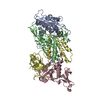
| ||||||||||||
|---|---|---|---|---|---|---|---|---|---|---|---|---|---|
| 1 |
| ||||||||||||
| Unit cell |
|
-Components
| #1: Protein | Mass: 22439.340 Da / Num. of mol.: 2 Source method: isolated from a genetically manipulated source Source: (gene. exp.) References: UniProt: J8G8J6, Hydrolases; Glycosylases; Hydrolysing N-glycosyl compounds #2: Protein | Mass: 22557.969 Da / Num. of mol.: 2 Source method: isolated from a genetically manipulated source Source: (gene. exp.) metagenome (others) / Production host: #3: Water | ChemComp-HOH / |  Mass: 18.015 Da / Num. of mol.: 767 / Source method: isolated from a natural source / Formula: H2O Mass: 18.015 Da / Num. of mol.: 767 / Source method: isolated from a natural source / Formula: H2OHas protein modification | N | |
|---|
-Experimental details
-Experiment
| Experiment | Method: X-RAY DIFFRACTION / Number of used crystals: 1 |
|---|
- Sample preparation
| Crystal | Density Matthews: 2.3 Å3/Da / Density % sol: 46.45 % |
|---|---|
| Crystal grow | Temperature: 291 K / Method: vapor diffusion, hanging drop / pH: 8 / Details: 0.1 M Tris pH 8.0 0.2 M MgCl2 25% PEG 3350 |
-Data collection
| Diffraction | Mean temperature: 80 K / Serial crystal experiment: N |
|---|---|
| Diffraction source | Source: SYNCHROTRON / Site: APS / Beamline: 24-ID-E / Wavelength: 0.9792 Å |
| Detector | Type: DECTRIS EIGER X 16M / Detector: PIXEL / Date: Dec 15, 2022 |
| Radiation | Protocol: SINGLE WAVELENGTH / Monochromatic (M) / Laue (L): M / Scattering type: x-ray |
| Radiation wavelength | Wavelength: 0.9792 Å / Relative weight: 1 |
| Reflection | Resolution: 1.8→35.63 Å / Num. obs: 75023 / % possible obs: 99.4 % / Redundancy: 4.3 % / Biso Wilson estimate: 26.51 Å2 / CC1/2: 0.998 / Net I/σ(I): 10 |
| Reflection shell | Resolution: 1.8→1.84 Å / Num. unique obs: 4316 / Rpim(I) all: 0.765 |
- Processing
| Software |
| |||||||||||||||||||||||||||||||||||||||||||||||||||||||||||||||||||||||||||||||||||||||||||||||||||||||||
|---|---|---|---|---|---|---|---|---|---|---|---|---|---|---|---|---|---|---|---|---|---|---|---|---|---|---|---|---|---|---|---|---|---|---|---|---|---|---|---|---|---|---|---|---|---|---|---|---|---|---|---|---|---|---|---|---|---|---|---|---|---|---|---|---|---|---|---|---|---|---|---|---|---|---|---|---|---|---|---|---|---|---|---|---|---|---|---|---|---|---|---|---|---|---|---|---|---|---|---|---|---|---|---|---|---|---|
| Refinement | Method to determine structure: MOLECULAR REPLACEMENT / Resolution: 1.8→35.63 Å / SU ML: 0.2047 / Cross valid method: FREE R-VALUE / σ(F): 1.34 / Phase error: 23.056 Stereochemistry target values: GeoStd + Monomer Library + CDL v1.2
| |||||||||||||||||||||||||||||||||||||||||||||||||||||||||||||||||||||||||||||||||||||||||||||||||||||||||
| Solvent computation | Shrinkage radii: 0.9 Å / VDW probe radii: 1.1 Å / Solvent model: FLAT BULK SOLVENT MODEL | |||||||||||||||||||||||||||||||||||||||||||||||||||||||||||||||||||||||||||||||||||||||||||||||||||||||||
| Displacement parameters | Biso mean: 33.92 Å2 | |||||||||||||||||||||||||||||||||||||||||||||||||||||||||||||||||||||||||||||||||||||||||||||||||||||||||
| Refinement step | Cycle: LAST / Resolution: 1.8→35.63 Å
| |||||||||||||||||||||||||||||||||||||||||||||||||||||||||||||||||||||||||||||||||||||||||||||||||||||||||
| Refine LS restraints |
| |||||||||||||||||||||||||||||||||||||||||||||||||||||||||||||||||||||||||||||||||||||||||||||||||||||||||
| LS refinement shell |
| |||||||||||||||||||||||||||||||||||||||||||||||||||||||||||||||||||||||||||||||||||||||||||||||||||||||||
| Refinement TLS params. | Method: refined / Origin x: -17.1766205307 Å / Origin y: -11.3157477284 Å / Origin z: -45.634775791 Å
| |||||||||||||||||||||||||||||||||||||||||||||||||||||||||||||||||||||||||||||||||||||||||||||||||||||||||
| Refinement TLS group | Selection details: all |
