[English] 日本語
Yorodumi
- PDB-9b4t: Crystal structure of the MRAS-p110alpha complex -

+
Open data


ID or keywords:

Loading...

-
Basic information

Entry
Database: PDB / ID: 9b4t
TitleCrystal structure of the MRAS-p110alpha complex
Components
  • Phosphatidylinositol 4,5-bisphosphate 3-kinase catalytic subunit alpha isoform
  • Ras-related protein M-Ras
KeywordsONCOPROTEIN / RAS / RRAS3 / MRAS / p110alpha / PIK3CA / PI3Kalpha / PI3Ka / PI3K
Function / homology
Function and homology information


protein phosphatase type 1 complex / response to muscle inactivity / negative regulation of actin filament depolymerization / GTP-dependent protein binding / response to L-leucine / regulation of actin filament organization / response to butyrate / IRS-mediated signalling / autosome genomic imprinting / phosphatidylinositol 3-kinase complex ...protein phosphatase type 1 complex / response to muscle inactivity / negative regulation of actin filament depolymerization / GTP-dependent protein binding / response to L-leucine / regulation of actin filament organization / response to butyrate / IRS-mediated signalling / autosome genomic imprinting / phosphatidylinositol 3-kinase complex / PI3K events in ERBB4 signaling / cellular response to hydrostatic pressure / regulation of cellular respiration / positive regulation of protein localization to membrane / Activated NTRK2 signals through PI3K / negative regulation of fibroblast apoptotic process / Activated NTRK3 signals through PI3K / phosphatidylinositol 3-kinase complex, class IB / SHOC2 M1731 mutant abolishes MRAS complex function / Gain-of-function MRAS complexes activate RAF signaling / vasculature development / 1-phosphatidylinositol-4-phosphate 3-kinase activity / Signaling by cytosolic FGFR1 fusion mutants / Co-stimulation by ICOS / cardiac muscle cell contraction / phosphatidylinositol 3-kinase complex, class IA / Nephrin family interactions / anoikis / Signaling by LTK in cancer / phosphatidylinositol-3-phosphate biosynthetic process / Signaling by LTK / MET activates PI3K/AKT signaling / PI3K/AKT activation / 1-phosphatidylinositol-4,5-bisphosphate 3-kinase activity / phosphatidylinositol-4,5-bisphosphate 3-kinase / phosphatidylinositol 3-kinase / vascular endothelial growth factor signaling pathway / relaxation of cardiac muscle / 1-phosphatidylinositol-3-kinase activity / Signaling by ALK / PI-3K cascade:FGFR3 / Erythropoietin activates Phosphoinositide-3-kinase (PI3K) / negative regulation of macroautophagy / PI-3K cascade:FGFR2 / phosphatidylinositol-mediated signaling / PI-3K cascade:FGFR4 / PI-3K cascade:FGFR1 / phosphatidylinositol phosphate biosynthetic process / Synthesis of PIPs at the plasma membrane / response to dexamethasone / negative regulation of anoikis / RET signaling / PI3K events in ERBB2 signaling / protein kinase activator activity / insulin receptor substrate binding / Interleukin-3, Interleukin-5 and GM-CSF signaling / PI3K Cascade / regulation of multicellular organism growth / intercalated disc / CD28 dependent PI3K/Akt signaling / positive regulation of TOR signaling / RAC2 GTPase cycle / Role of LAT2/NTAL/LAB on calcium mobilization / Interleukin receptor SHC signaling / GAB1 signalosome / Role of phospholipids in phagocytosis / adipose tissue development / phagocytosis / endothelial cell migration / Signaling by PDGFRA transmembrane, juxtamembrane and kinase domain mutants / Signaling by PDGFRA extracellular domain mutants / Signaling by FGFR4 in disease / positive regulation of lamellipodium assembly / energy homeostasis / Signaling by FLT3 ITD and TKD mutants / cardiac muscle contraction / GPVI-mediated activation cascade / Signaling by FGFR3 in disease / Tie2 Signaling / Signaling by FGFR2 in disease / response to muscle stretch / T cell costimulation / RAC1 GTPase cycle / Signaling by FLT3 fusion proteins / FLT3 Signaling / Signaling by FGFR1 in disease / Downstream signal transduction / insulin-like growth factor receptor signaling pathway / cellular response to leukemia inhibitory factor / Signaling by phosphorylated juxtamembrane, extracellular and kinase domain KIT mutants / liver development / small monomeric GTPase / response to activity / phosphatidylinositol 3-kinase/protein kinase B signal transduction / Regulation of signaling by CBL / positive regulation of smooth muscle cell proliferation / RAF activation / cellular response to glucose stimulus / Signaling by SCF-KIT / Constitutive Signaling by EGFRvIII
Similarity search - Function
PI3Kalpha, catalytic domain / PI3-kinase family, p85-binding domain / PI3-kinase family, p85-binding domain / Phosphatidylinositol 3-kinase, adaptor-binding domain / Phosphatidylinositol 3-kinase adaptor-binding (PI3K ABD) domain profile. / PI3-kinase family, Ras-binding domain / Phosphatidylinositol 3-kinase Ras-binding (PI3K RBD) domain / PI3-kinase family, ras-binding domain / Phosphatidylinositol 3-kinase Ras-binding (PI3K RBD) domain profile. / C2 phosphatidylinositol 3-kinase-type domain ...PI3Kalpha, catalytic domain / PI3-kinase family, p85-binding domain / PI3-kinase family, p85-binding domain / Phosphatidylinositol 3-kinase, adaptor-binding domain / Phosphatidylinositol 3-kinase adaptor-binding (PI3K ABD) domain profile. / PI3-kinase family, Ras-binding domain / Phosphatidylinositol 3-kinase Ras-binding (PI3K RBD) domain / PI3-kinase family, ras-binding domain / Phosphatidylinositol 3-kinase Ras-binding (PI3K RBD) domain profile. / C2 phosphatidylinositol 3-kinase-type domain / Phosphoinositide 3-kinase C2 / C2 phosphatidylinositol 3-kinase (PI3K)-type domain profile. / Phosphoinositide 3-kinase, region postulated to contain C2 domain / Phosphoinositide 3-kinase family, accessory domain (PIK domain) / Phosphoinositide 3-kinase family, accessory domain (PIK domain) / Phosphoinositide 3-kinase, accessory (PIK) domain superfamily / Phosphoinositide 3-kinase, accessory (PIK) domain / Phosphatidylinositol kinase / PIK helical domain profile. / Phosphatidylinositol 3- and 4-kinases signature 1. / Phosphatidylinositol 3/4-kinase, conserved site / Phosphatidylinositol 3- and 4-kinases signature 2. / Small GTPase, Ras-type / Phosphatidylinositol 3-/4-kinase, catalytic domain superfamily / small GTPase Ras family profile. / Phosphoinositide 3-kinase, catalytic domain / Phosphatidylinositol 3- and 4-kinase / Phosphatidylinositol 3- and 4-kinases catalytic domain profile. / Phosphatidylinositol 3-/4-kinase, catalytic domain / Ran (Ras-related nuclear proteins) /TC4 subfamily of small GTPases / C2 domain superfamily / Rho (Ras homology) subfamily of Ras-like small GTPases / Ras subfamily of RAS small GTPases / Small GTPase / Ras family / Rab subfamily of small GTPases / Small GTP-binding protein domain / Armadillo-type fold / Ubiquitin-like domain superfamily / Protein kinase-like domain superfamily / P-loop containing nucleoside triphosphate hydrolase
Similarity search - Domain/homology
Chem-5H5 / PHOSPHOAMINOPHOSPHONIC ACID-GUANYLATE ESTER / Ras-related protein M-Ras / Phosphatidylinositol 4,5-bisphosphate 3-kinase catalytic subunit alpha isoform
Similarity search - Component
Biological speciesHomo sapiens (human)
MethodX-RAY DIFFRACTION / SYNCHROTRON / MOLECULAR REPLACEMENT / Resolution: 2.75 Å
AuthorsCzyzyk, D.J. / Simanshu, D.K.
Funding support United States, 1items
OrganizationGrant numberCountry
National Institutes of Health/National Cancer Institute (NIH/NCI)75N91019D00024 United States
CitationJournal: Nat Commun / Year: 2025
Title: Structural insights into isoform-specific RAS-PI3K alpha interactions and the role of RAS in PI3K alpha activation.
Authors: Czyzyk, D. / Yan, W. / Messing, S. / Gillette, W. / Tsuji, T. / Yamaguchi, M. / Furuzono, S. / Turner, D.M. / Esposito, D. / Nissley, D.V. / McCormick, F. / Simanshu, D.K.
History
DepositionMar 21, 2024Deposition site: RCSB / Processing site: RCSB
Revision 1.0Jan 22, 2025Provider: repository / Type: Initial release

-
Structure visualization

Structure viewerMolecule:
MolmilJmol/JSmol

Downloads & links

-
Assembly

Deposited unit
A: Phosphatidylinositol 4,5-bisphosphate 3-kinase catalytic subunit alpha isoform
B: Ras-related protein M-Ras
hetero molecules


Theoretical massNumber of molelcules
Total (without water)133,3475
Polymers132,4182
Non-polymers9293
Water48627
1


  • Idetical with deposited unit
  • defined by author&software
  • Evidence: gel filtration
TypeNameSymmetry operationNumber
identity operation1_555x,y,z1
Buried area2800 Å2
ΔGint-18 kcal/mol
Surface area47020 Å2
MethodPISA
Unit cell
Length a, b, c (Å)58.935, 105.936, 114.493
Angle α, β, γ (deg.)90.00, 97.50, 90.00
Int Tables number4
Space group name H-MP1211

-
Components

-
Protein , 2 types, 2 molecules AB

#1: Protein Phosphatidylinositol 4,5-bisphosphate 3-kinase catalytic subunit alpha isoform / PI3-kinase subunit alpha / PI3K-alpha / PI3Kalpha / PtdIns-3-kinase subunit alpha / ...PI3-kinase subunit alpha / PI3K-alpha / PI3Kalpha / PtdIns-3-kinase subunit alpha / Phosphatidylinositol 4 / 5-bisphosphate 3-kinase 110 kDa catalytic subunit alpha / PtdIns-3-kinase subunit p110-alpha / p110alpha / Phosphoinositide 3-kinase alpha / Phosphoinositide-3-kinase catalytic alpha polypeptide / Serine/threonine protein kinase PIK3CA


Mass: 112033.430 Da / Num. of mol.: 1 / Mutation: W1057A, I1058A, F1059A
Source method: isolated from a genetically manipulated source
Source: (gene. exp.) Homo sapiens (human) / Gene: PIK3CA / Production host: Trichoplusia (butterflies/moths)
References: UniProt: P42336, phosphatidylinositol 3-kinase, phosphatidylinositol-4,5-bisphosphate 3-kinase, non-specific serine/threonine protein kinase
#2: Protein Ras-related protein M-Ras / Ras-related protein R-Ras3


Mass: 20384.250 Da / Num. of mol.: 1 / Mutation: Q35A
Source method: isolated from a genetically manipulated source
Source: (gene. exp.) Homo sapiens (human) / Gene: MRAS, RRAS3 / Production host: Escherichia coli (E. coli) / References: UniProt: O14807, small monomeric GTPase

-
Non-polymers , 4 types, 30 molecules

#3: Chemical ChemComp-5H5 / (2S)-2-({2-[1-(propan-2-yl)-1H-1,2,4-triazol-5-yl]-5,6-dihydroimidazo[1,2-d][1,4]benzoxazepin-9-yl}oxy)propanamide / GDC-0326


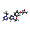
Mass: 382.416 Da / Num. of mol.: 1 / Source method: obtained synthetically / Formula: C19H22N6O3 / Feature type: SUBJECT OF INVESTIGATION
#4: Chemical ChemComp-GNP / PHOSPHOAMINOPHOSPHONIC ACID-GUANYLATE ESTER


Mass: 522.196 Da / Num. of mol.: 1 / Source method: obtained synthetically / Formula: C10H17N6O13P3 / Feature type: SUBJECT OF INVESTIGATION
Comment: GppNHp, GMPPNP, energy-carrying molecule analogue*YM
#5: Chemical ChemComp-MG / MAGNESIUM ION


Mass: 24.305 Da / Num. of mol.: 1 / Source method: obtained synthetically / Formula: Mg
#6: Water ChemComp-HOH / water


Mass: 18.015 Da / Num. of mol.: 27 / Source method: isolated from a natural source / Formula: H2O

-
Details

Has ligand of interestY
Has protein modificationN

-
Experimental details

-
Experiment

ExperimentMethod: X-RAY DIFFRACTION / Number of used crystals: 1

-
Sample preparation

CrystalDensity Matthews: 2.67 Å3/Da / Density % sol: 53.89 %
Crystal growTemperature: 293 K / Method: vapor diffusion, sitting drop / pH: 8.5
Details: 0.1 M (Tris/Bicine) 1.2%w/v cholic acid mix, 50%v/v (40%v/v ethylene glycol, 20%w/v PEG 8000)

-
Data collection

DiffractionMean temperature: 100 K / Serial crystal experiment: N
Diffraction sourceSource: SYNCHROTRON / Site: APS / Beamline: 24-ID-E / Wavelength: 0.97918 Å
DetectorType: DECTRIS EIGER X 16M / Detector: PIXEL / Date: Dec 17, 2022
RadiationProtocol: SINGLE WAVELENGTH / Monochromatic (M) / Laue (L): M / Scattering type: x-ray
Radiation wavelengthWavelength: 0.97918 Å / Relative weight: 1
ReflectionResolution: 2.75→50.03 Å / Num. obs: 35877 / % possible obs: 98.7 % / Redundancy: 7 % / CC1/2: 0.999 / Rmerge(I) obs: 0.078 / Rrim(I) all: 0.084 / Net I/σ(I): 13.48
Reflection shell
Resolution (Å)Rmerge(I) obsNum. unique obsCC1/2Rrim(I) allDiffraction-ID
2.75-2.921.11957130.871.2051
2.92-3.120.61154480.9590.6581
3.12-3.370.3450550.9810.3681
3.37-3.690.1846230.9920.1941
3.69-4.120.10142150.9970.1091
4.12-4.750.06937520.9980.0751
4.75-5.810.05931670.9980.0641
5.81-8.160.05125000.9980.0551
8.16-50.030.03414040.9990.0371

-
Processing

Software
NameVersionClassification
PHENIX(1.21_5207: ???)refinement
XDSdata scaling
XDSdata reduction
PHASERphasing
RefinementMethod to determine structure: MOLECULAR REPLACEMENT / Resolution: 2.75→50.03 Å / SU ML: 0.5 / Cross valid method: FREE R-VALUE / σ(F): 1.35 / Phase error: 34.13 / Stereochemistry target values: ML
RfactorNum. reflection% reflection
Rfree0.2619 1787 5 %
Rwork0.2135 --
obs0.2158 35732 98.31 %
Solvent computationShrinkage radii: 0.9 Å / VDW probe radii: 1.1 Å / Solvent model: FLAT BULK SOLVENT MODEL
Refinement stepCycle: LAST / Resolution: 2.75→50.03 Å
ProteinNucleic acidLigandSolventTotal
Num. atoms8626 0 61 27 8714
Refine LS restraints
Refine-IDTypeDev idealNumber
X-RAY DIFFRACTIONf_bond_d0.0028878
X-RAY DIFFRACTIONf_angle_d0.40612014
X-RAY DIFFRACTIONf_dihedral_angle_d11.6813366
X-RAY DIFFRACTIONf_chiral_restr0.0381317
X-RAY DIFFRACTIONf_plane_restr0.0031533
LS refinement shell
Resolution (Å)Rfactor RfreeNum. reflection RfreeRfactor RworkNum. reflection RworkRefine-ID% reflection obs (%)
2.75-2.830.49951360.45472582X-RAY DIFFRACTION96
2.83-2.910.39421360.37572554X-RAY DIFFRACTION98
2.91-30.32961350.32112604X-RAY DIFFRACTION98
3-3.110.33781370.28462619X-RAY DIFFRACTION98
3.11-3.230.30431370.27772586X-RAY DIFFRACTION98
3.23-3.380.3281350.28562580X-RAY DIFFRACTION98
3.38-3.560.34131350.25772557X-RAY DIFFRACTION97
3.56-3.780.30271390.2252625X-RAY DIFFRACTION99
3.78-4.080.22961380.20982635X-RAY DIFFRACTION99
4.08-4.490.24981400.19822641X-RAY DIFFRACTION99
4.49-5.130.22841360.17672595X-RAY DIFFRACTION98
5.13-6.460.28781420.20812682X-RAY DIFFRACTION100
6.47-50.030.19531410.16812685X-RAY DIFFRACTION99
Refinement TLS params.Method: refined / Origin x: -1.6934 Å / Origin y: -12.2223 Å / Origin z: 37.8298 Å
111213212223313233
T0.6517 Å20.0024 Å2-0.0368 Å2-0.8096 Å2-0.0261 Å2--0.6542 Å2
L0.8789 °2-0.2281 °2-0.2751 °2-1.0712 °2-0.1974 °2--1.7841 °2
S0.0273 Å °0.0975 Å °-0.0197 Å °0.0458 Å °0.0764 Å °0.051 Å °-0.204 Å °0.133 Å °-0.1024 Å °
Refinement TLS groupSelection details: all

+
About Yorodumi

-
News

-
Feb 9, 2022. New format data for meta-information of EMDB entries

New format data for meta-information of EMDB entries

  • Version 3 of the EMDB header file is now the official format.
  • The previous official version 1.9 will be removed from the archive.

Related info.:EMDB header

External links:wwPDB to switch to version 3 of the EMDB data model

-
Aug 12, 2020. Covid-19 info

Covid-19 info

URL: https://pdbj.org/emnavi/covid19.php

New page: Covid-19 featured information page in EM Navigator.

Related info.:Covid-19 info / Mar 5, 2020. Novel coronavirus structure data

+
Mar 5, 2020. Novel coronavirus structure data

Novel coronavirus structure data

Related info.:Yorodumi Speices / Aug 12, 2020. Covid-19 info

External links:COVID-19 featured content - PDBj / Molecule of the Month (242):Coronavirus Proteases

+
Jan 31, 2019. EMDB accession codes are about to change! (news from PDBe EMDB page)

EMDB accession codes are about to change! (news from PDBe EMDB page)

  • The allocation of 4 digits for EMDB accession codes will soon come to an end. Whilst these codes will remain in use, new EMDB accession codes will include an additional digit and will expand incrementally as the available range of codes is exhausted. The current 4-digit format prefixed with “EMD-” (i.e. EMD-XXXX) will advance to a 5-digit format (i.e. EMD-XXXXX), and so on. It is currently estimated that the 4-digit codes will be depleted around Spring 2019, at which point the 5-digit format will come into force.
  • The EM Navigator/Yorodumi systems omit the EMD- prefix.

Related info.:Q: What is EMD? / ID/Accession-code notation in Yorodumi/EM Navigator

External links:EMDB Accession Codes are Changing Soon! / Contact to PDBj

+
Jul 12, 2017. Major update of PDB

Major update of PDB

  • wwPDB released updated PDB data conforming to the new PDBx/mmCIF dictionary.
  • This is a major update changing the version number from 4 to 5, and with Remediation, in which all the entries are updated.
  • In this update, many items about electron microscopy experimental information are reorganized (e.g. em_software).
  • Now, EM Navigator and Yorodumi are based on the updated data.

External links:wwPDB Remediation / Enriched Model Files Conforming to OneDep Data Standards Now Available in the PDB FTP Archive

-
Yorodumi

Thousand views of thousand structures

  • Yorodumi is a browser for structure data from EMDB, PDB, SASBDB, etc.
  • This page is also the successor to EM Navigator detail page, and also detail information page/front-end page for Omokage search.
  • The word "yorodu" (or yorozu) is an old Japanese word meaning "ten thousand". "mi" (miru) is to see.

Related info.:EMDB / PDB / SASBDB / Comparison of 3 databanks / Yorodumi Search / Aug 31, 2016. New EM Navigator & Yorodumi / Yorodumi Papers / Jmol/JSmol / Function and homology information / Changes in new EM Navigator and Yorodumi

Read more