 Movie
Movie Controller


+ Open data

- Basic information
| Entry | Database: PDB / ID: 9ayi | ||||||
|---|---|---|---|---|---|---|---|
| Title | Human malic enzyme 2 cofactor complex at 1.89 Angstrom. | ||||||
 Components Components | NAD-dependent malic enzyme, mitochondrial | ||||||
 Keywords Keywords | OXIDOREDUCTASE / Malic enzyme 2 / NAD+ / co-factor complex / ME2 / mitochondrial localisation / m-NAD-ME | ||||||
| Function / homology |  Function and homology information Function and homology informationmalate dehydrogenase (oxaloacetate-decarboxylating) / regulation of NADP metabolic process / malate dehydrogenase (decarboxylating) (NAD+) activity / malic enzyme activity / malate dehydrogenase (decarboxylating) (NADP+) activity / oxaloacetate decarboxylase activity / malate metabolic process / pyruvate metabolic process / Pyruvate metabolism / Mitochondrial protein degradation ...malate dehydrogenase (oxaloacetate-decarboxylating) / regulation of NADP metabolic process / malate dehydrogenase (decarboxylating) (NAD+) activity / malic enzyme activity / malate dehydrogenase (decarboxylating) (NADP+) activity / oxaloacetate decarboxylase activity / malate metabolic process / pyruvate metabolic process / Pyruvate metabolism / Mitochondrial protein degradation / NAD binding / electron transfer activity / mitochondrial matrix / intracellular membrane-bounded organelle / mitochondrion / metal ion binding Similarity search - Function | ||||||
| Biological species |  Homo sapiens (human) Homo sapiens (human) | ||||||
| Method |  X-RAY DIFFRACTION / SYNCHROTRON / MOLECULAR REPLACEMENT / Resolution: 1.89 Å X-RAY DIFFRACTION / SYNCHROTRON / MOLECULAR REPLACEMENT / Resolution: 1.89 Å | ||||||
 Authors Authors | Krinkel, B.A. / Squire, C.J. / Loomes, K.M. | ||||||
| Funding support | 1items
| ||||||
 Citation Citation | Journal: To Be Published Title: Human malic enzyme 2 Authors: Krinkel, B.A. / Squire, C.J. / Loomes, K.M. | ||||||
| History |
|
- Structure visualization
| Structure viewer | Molecule: MolmilJmol/JSmol |
|---|
- Downloads & links
-Download
| PDBx/mmCIF format | 9ayi.cif.gz | 494.5 KB | Display | PDBx/mmCIF format |
|---|---|---|---|---|
| PDB format | pdb9ayi.ent.gz | 401.2 KB | Display | PDB format |
| PDBx/mmJSON format | 9ayi.json.gz | Tree view | PDBx/mmJSON format | |
| Others |  Other downloads Other downloads |
-Validation report
| Summary document | 9ayi_validation.pdf.gz | 3.5 MB | Display | wwPDB validaton report |
|---|---|---|---|---|
| Full document | 9ayi_full_validation.pdf.gz | 3.5 MB | Display | |
| Data in XML | 9ayi_validation.xml.gz | 115 KB | Display | |
| Data in CIF | 9ayi_validation.cif.gz | 156.1 KB | Display | |
| Arichive directory | https://data.pdbj.org/pub/pdb/validation_reports/ay/9ayiftp://data.pdbj.org/pub/pdb/validation_reports/ay/9ayi | HTTPS FTP |
-Related structure data
| Related structure data |  8w24 |
|---|---|
| Similar structure data |
-Links
 PDBj
PDBj
- Assembly
| Deposited unit | 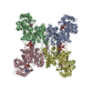
| ||||||||
|---|---|---|---|---|---|---|---|---|---|
| 1 |
| ||||||||
| Unit cell |
|
-Components
-Protein , 1 types, 4 molecules ABCD
| #1: Protein | Mass: 64706.309 Da / Num. of mol.: 4 Source method: isolated from a genetically manipulated source Source: (gene. exp.) Homo sapiens (human) / Gene: ME2 / Production host:  References: UniProt: P23368, malate dehydrogenase (oxaloacetate-decarboxylating) |
|---|
-Non-polymers , 6 types, 1983 molecules 


| #2: Chemical | ChemComp-NAD /  Mass: 663.425 Da / Num. of mol.: 8 / Source method: obtained synthetically / Formula: C21H27N7O14P2 / Comment: NAD*YM Mass: 663.425 Da / Num. of mol.: 8 / Source method: obtained synthetically / Formula: C21H27N7O14P2 / Comment: NAD*YM#3: Chemical | ChemComp-EDO /  Mass: 62.068 Da / Num. of mol.: 5 / Source method: obtained synthetically / Formula: C2H6O2 Mass: 62.068 Da / Num. of mol.: 5 / Source method: obtained synthetically / Formula: C2H6O2#4: Chemical | ChemComp-CL /  Mass: 35.453 Da / Num. of mol.: 4 / Source method: obtained synthetically / Formula: Cl Mass: 35.453 Da / Num. of mol.: 4 / Source method: obtained synthetically / Formula: Cl#5: Chemical |  Mass: 24.305 Da / Num. of mol.: 2 / Source method: obtained synthetically / Formula: Mg Mass: 24.305 Da / Num. of mol.: 2 / Source method: obtained synthetically / Formula: Mg#6: Chemical | ChemComp-TLA /  Mass: 150.087 Da / Num. of mol.: 4 / Source method: obtained synthetically / Formula: C4H6O6 Mass: 150.087 Da / Num. of mol.: 4 / Source method: obtained synthetically / Formula: C4H6O6#7: Water | ChemComp-HOH / | Mass: 18.015 Da / Num. of mol.: 1960 / Source method: isolated from a natural source / Formula: H2O |
|---|
-Details
| Has ligand of interest | N |
|---|---|
| Has protein modification | Y |
-Experimental details
-Experiment
| Experiment | Method: X-RAY DIFFRACTION / Number of used crystals: 1 |
|---|
- Sample preparation
| Crystal | Density Matthews: 2.67 Å3/Da / Density % sol: 53.87 % |
|---|---|
| Crystal grow | Temperature: 291 K / Method: vapor diffusion, sitting drop / pH: 7 Details: 0.1 M Bis-Tris, 0.1 M Ammonium Tartrate, 0.02 M Potassium Citrate, 20% PEG-3350. |
-Data collection
| Diffraction | Mean temperature: 100 K / Serial crystal experiment: N |
|---|---|
| Diffraction source | Source: SYNCHROTRON / Site: Australian Synchrotron  / Beamline: MX2 / Wavelength: 0.9537 Å / Beamline: MX2 / Wavelength: 0.9537 Å |
| Detector | Type: DECTRIS EIGER X 16M / Detector: PIXEL / Date: Jul 7, 2023 |
| Radiation | Protocol: SINGLE WAVELENGTH / Monochromatic (M) / Laue (L): M / Scattering type: x-ray |
| Radiation wavelength | Wavelength: 0.9537 Å / Relative weight: 1 |
| Reflection | Resolution: 1.89→47.42 Å / Num. obs: 200119 / % possible obs: 98.2 % / Redundancy: 3.6 % / CC1/2: 0.99 / Net I/σ(I): 6.9 |
| Reflection shell | Resolution: 1.89→1.93 Å / Redundancy: 3.5 % / Mean I/σ(I) obs: 1.6 / Num. unique obs: 8176 / CC1/2: 0.483 / % possible all: 79.9 |
- Processing
| Software |
| ||||||||||||||||||||||||||||||||||||||||||||||||||||||||||||||||||||||||||||||||||||||||||||||||||||||||||||||||||||||||||||||||||||||||||||||||||||||||||||||||||||||||||||||||||||||
|---|---|---|---|---|---|---|---|---|---|---|---|---|---|---|---|---|---|---|---|---|---|---|---|---|---|---|---|---|---|---|---|---|---|---|---|---|---|---|---|---|---|---|---|---|---|---|---|---|---|---|---|---|---|---|---|---|---|---|---|---|---|---|---|---|---|---|---|---|---|---|---|---|---|---|---|---|---|---|---|---|---|---|---|---|---|---|---|---|---|---|---|---|---|---|---|---|---|---|---|---|---|---|---|---|---|---|---|---|---|---|---|---|---|---|---|---|---|---|---|---|---|---|---|---|---|---|---|---|---|---|---|---|---|---|---|---|---|---|---|---|---|---|---|---|---|---|---|---|---|---|---|---|---|---|---|---|---|---|---|---|---|---|---|---|---|---|---|---|---|---|---|---|---|---|---|---|---|---|---|---|---|---|---|
| Refinement | Method to determine structure: MOLECULAR REPLACEMENT / Resolution: 1.89→47.42 Å / Cor.coef. Fo:Fc: 0.962 / Cor.coef. Fo:Fc free: 0.954 / SU B: 3.54 / SU ML: 0.1 / Cross valid method: THROUGHOUT / ESU R: 0.148 / ESU R Free: 0.127 / Stereochemistry target values: MAXIMUM LIKELIHOOD / Details: HYDROGENS HAVE BEEN ADDED IN THE RIDING POSITIONS
| ||||||||||||||||||||||||||||||||||||||||||||||||||||||||||||||||||||||||||||||||||||||||||||||||||||||||||||||||||||||||||||||||||||||||||||||||||||||||||||||||||||||||||||||||||||||
| Solvent computation | Ion probe radii: 0.8 Å / Shrinkage radii: 0.8 Å / VDW probe radii: 1.2 Å / Solvent model: MASK | ||||||||||||||||||||||||||||||||||||||||||||||||||||||||||||||||||||||||||||||||||||||||||||||||||||||||||||||||||||||||||||||||||||||||||||||||||||||||||||||||||||||||||||||||||||||
| Displacement parameters | Biso mean: 29.068 Å2
| ||||||||||||||||||||||||||||||||||||||||||||||||||||||||||||||||||||||||||||||||||||||||||||||||||||||||||||||||||||||||||||||||||||||||||||||||||||||||||||||||||||||||||||||||||||||
| Refinement step | Cycle: 1 / Resolution: 1.89→47.42 Å
| ||||||||||||||||||||||||||||||||||||||||||||||||||||||||||||||||||||||||||||||||||||||||||||||||||||||||||||||||||||||||||||||||||||||||||||||||||||||||||||||||||||||||||||||||||||||
| Refine LS restraints |
|