 Movie
Movie Controller


+ Open data

- Basic information
| Entry | Database: PDB / ID: 9av0 | ||||||
|---|---|---|---|---|---|---|---|
| Title | Crystal structure of S. aureus GuaB dCBS with inhibitor GNE2011 | ||||||
 Components Components | Inosine-5'-monophosphate dehydrogenase | ||||||
 Keywords Keywords | OXIDOREDUCTASE / GuaB / inhibitor | ||||||
| Function / homology |  Function and homology information Function and homology informationIMP dehydrogenase / IMP dehydrogenase activity / GMP biosynthetic process / GTP biosynthetic process / nucleotide binding / metal ion binding Similarity search - Function | ||||||
| Biological species |   Staphylococcus aureus (bacteria) Staphylococcus aureus (bacteria) | ||||||
| Method |  X-RAY DIFFRACTION / SYNCHROTRON / MOLECULAR REPLACEMENT / Resolution: 2.2 Å X-RAY DIFFRACTION / SYNCHROTRON / MOLECULAR REPLACEMENT / Resolution: 2.2 Å | ||||||
 Authors Authors | Harris, S.F. / Wu, P. | ||||||
| Funding support | 1items
| ||||||
 Citation Citation | Journal: Mbio / Year: 2024 Title: Discovery of GuaB inhibitors with efficacy against Acinetobacter baumannii infection. Authors: Kofoed, E.M. / Aliagas, I. / Crawford, T. / Mao, J. / Harris, S.F. / Xu, M. / Wang, S. / Wu, P. / Ma, F. / Clark, K. / Sims, J. / Xu, Y. / Peng, Y. / Skippington, E. / Yang, Y. / Reeder, J. ...Authors: Kofoed, E.M. / Aliagas, I. / Crawford, T. / Mao, J. / Harris, S.F. / Xu, M. / Wang, S. / Wu, P. / Ma, F. / Clark, K. / Sims, J. / Xu, Y. / Peng, Y. / Skippington, E. / Yang, Y. / Reeder, J. / Ubhayakar, S. / Baumgardner, M. / Yan, Z. / Chen, J. / Park, S. / Zhang, H. / Yen, C.-W. / Lorenzo, M. / Skelton, N. / Liang, X. / Chen, L. / Hoag, B. / Li, C.S. / Liu, Z. / Wai, J. / Liu, X. / Liang, J. / Tan, M.W. #1: Journal: Bioorg.Med.Chem.Lett. / Year: 2024Title: Discovery of potent dihydro-oxazinoquinolinone inhibitors of GuaB for the treatment of tuberculosis. Authors: Zhou, Y. / Aliagas, I. / Wang, S. / Li, C.S. / Liu, Z. / Bowman, C.M. / Burdick, D.J. / Clark, K.R. / Dening, T.J. / Flygare, J. / Ganti, A. / Girgis, H.S. / Hanan, E.J. / Harris, S.F. / Hu, ...Authors: Zhou, Y. / Aliagas, I. / Wang, S. / Li, C.S. / Liu, Z. / Bowman, C.M. / Burdick, D.J. / Clark, K.R. / Dening, T.J. / Flygare, J. / Ganti, A. / Girgis, H.S. / Hanan, E.J. / Harris, S.F. / Hu, C. / Kapadia, S.B. / Koehler, M.F.T. / Lai, T. / Liang, J. / Liu, X. / Ma, F. / Mao, J. / Nicolai, J. / Sims, J. / Unhayaker, S. / Wai, J. / Wang, X. / Wu, P. / Xu, Y. / Yen, C.W. / Zhang, R. / Elfert, T.F. / Tan, M.W. / Kofoed, E.M. / Crawford, T.D. | ||||||
| History |
|
- Structure visualization
| Structure viewer | Molecule: MolmilJmol/JSmol |
|---|
- Downloads & links
-Download
| PDBx/mmCIF format | 9av0.cif.gz | 84 KB | Display | PDBx/mmCIF format |
|---|---|---|---|---|
| PDB format | pdb9av0.ent.gz | Display | PDB format | |
| PDBx/mmJSON format | 9av0.json.gz | Tree view | PDBx/mmJSON format | |
| Others |  Other downloads Other downloads |
-Validation report
| Summary document | 9av0_validation.pdf.gz | 1011.4 KB | Display | wwPDB validaton report |
|---|---|---|---|---|
| Full document | 9av0_full_validation.pdf.gz | 1015.1 KB | Display | |
| Data in XML | 9av0_validation.xml.gz | 15.8 KB | Display | |
| Data in CIF | 9av0_validation.cif.gz | 20.4 KB | Display | |
| Arichive directory | https://data.pdbj.org/pub/pdb/validation_reports/av/9av0ftp://data.pdbj.org/pub/pdb/validation_reports/av/9av0 | HTTPS FTP |
-Related structure data
| Related structure data |  9auvC  9auwC  9auxC 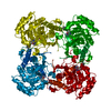 9auyC  9auzC  9av1C 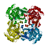 9av2C  9av3C C: citing same article ( |
|---|---|
| Similar structure data |
-Links
 PDBj
PDBj
- Assembly
| Deposited unit | 
| ||||||||
|---|---|---|---|---|---|---|---|---|---|
| 1 | 
| ||||||||
| Unit cell |
|
-Components
| #1: Protein | Mass: 40598.129 Da / Num. of mol.: 1 Source method: isolated from a genetically manipulated source Source: (gene. exp.) Staphylococcus aureus (bacteria) / Gene: guaB, SAUSA300_0388 / Production host: |
|---|---|
| #2: Chemical | ChemComp-IMP /   Mass: 348.206 Da / Num. of mol.: 1 / Source method: obtained synthetically / Formula: C10H13N4O8P Mass: 348.206 Da / Num. of mol.: 1 / Source method: obtained synthetically / Formula: C10H13N4O8P |
| #3: Chemical | ChemComp-A1AG0 / Mass: 406.865 Da / Num. of mol.: 1 / Source method: obtained synthetically / Formula: C22H19ClN4O2 / Feature type: SUBJECT OF INVESTIGATION |
| #4: Water | ChemComp-HOH /  Mass: 18.015 Da / Num. of mol.: 29 / Source method: isolated from a natural source / Formula: H2O Mass: 18.015 Da / Num. of mol.: 29 / Source method: isolated from a natural source / Formula: H2O |
| Has ligand of interest | Y |
| Has protein modification | N |
-Experimental details
-Experiment
| Experiment | Method: X-RAY DIFFRACTION / Number of used crystals: 1 |
|---|
- Sample preparation
| Crystal | Density Matthews: 2.16 Å3/Da / Density % sol: 43.14 % |
|---|---|
| Crystal grow | Temperature: 293 K / Method: vapor diffusion, sitting drop / pH: 9 / Details: 0.1M Bicine pH 9, 0.1M NaCl, 30% PEG550 MME |
-Data collection
| Diffraction | Mean temperature: 100 K / Serial crystal experiment: N |
|---|---|
| Diffraction source | Source: SYNCHROTRON / Site: APS  / Beamline: 24-ID-C / Wavelength: 0.9792 Å / Beamline: 24-ID-C / Wavelength: 0.9792 Å |
| Detector | Type: DECTRIS PILATUS 6M / Detector: PIXEL / Date: Feb 28, 2019 |
| Radiation | Protocol: SINGLE WAVELENGTH / Monochromatic (M) / Laue (L): M / Scattering type: x-ray |
| Radiation wavelength | Wavelength: 0.9792 Å / Relative weight: 1 |
| Reflection | Resolution: 2.2→73.85 Å / Num. obs: 17517 / % possible obs: 98.4 % / Redundancy: 6.6 % / CC1/2: 0.999 / Rmerge(I) obs: 0.055 / Rpim(I) all: 0.023 / Rrim(I) all: 0.06 / Net I/σ(I): 16.6 / Num. measured all: 115512 |
| Reflection shell | Resolution: 2.2→2.32 Å / % possible obs: 90.7 % / Redundancy: 6.7 % / Rmerge(I) obs: 0.862 / Num. measured all: 15795 / Num. unique obs: 2351 / CC1/2: 0.719 / Rpim(I) all: 0.357 / Rrim(I) all: 0.934 / Net I/σ(I) obs: 1.9 |
- Processing
| Software |
| ||||||||||||||||||||||||||||||||||||||||||||||||||||||||||||||||||||||||||||||||||||||||||||||||||||||||||||||||||
|---|---|---|---|---|---|---|---|---|---|---|---|---|---|---|---|---|---|---|---|---|---|---|---|---|---|---|---|---|---|---|---|---|---|---|---|---|---|---|---|---|---|---|---|---|---|---|---|---|---|---|---|---|---|---|---|---|---|---|---|---|---|---|---|---|---|---|---|---|---|---|---|---|---|---|---|---|---|---|---|---|---|---|---|---|---|---|---|---|---|---|---|---|---|---|---|---|---|---|---|---|---|---|---|---|---|---|---|---|---|---|---|---|---|---|---|
| Refinement | Method to determine structure: MOLECULAR REPLACEMENT / Resolution: 2.2→73.85 Å / Cor.coef. Fo:Fc: 0.938 / Cor.coef. Fo:Fc free: 0.928 / Rfactor Rfree error: 0 / SU R Cruickshank DPI: 0.302 / Cross valid method: THROUGHOUT / σ(F): 0 / SU R Blow DPI: 0.287 / SU Rfree Blow DPI: 0.207 / SU Rfree Cruickshank DPI: 0.214
| ||||||||||||||||||||||||||||||||||||||||||||||||||||||||||||||||||||||||||||||||||||||||||||||||||||||||||||||||||
| Displacement parameters | Biso mean: 76.29 Å2
| ||||||||||||||||||||||||||||||||||||||||||||||||||||||||||||||||||||||||||||||||||||||||||||||||||||||||||||||||||
| Refine analyze | Luzzati coordinate error obs: 0.41 Å | ||||||||||||||||||||||||||||||||||||||||||||||||||||||||||||||||||||||||||||||||||||||||||||||||||||||||||||||||||
| Refinement step | Cycle: 1 / Resolution: 2.2→73.85 Å
| ||||||||||||||||||||||||||||||||||||||||||||||||||||||||||||||||||||||||||||||||||||||||||||||||||||||||||||||||||
| Refine LS restraints |
| ||||||||||||||||||||||||||||||||||||||||||||||||||||||||||||||||||||||||||||||||||||||||||||||||||||||||||||||||||
| LS refinement shell | Resolution: 2.2→2.33 Å / Rfactor Rfree error: 0 / Total num. of bins used: 9
|
